HEALTH MONITORING: ARTHROPODS
ARACHNIDS
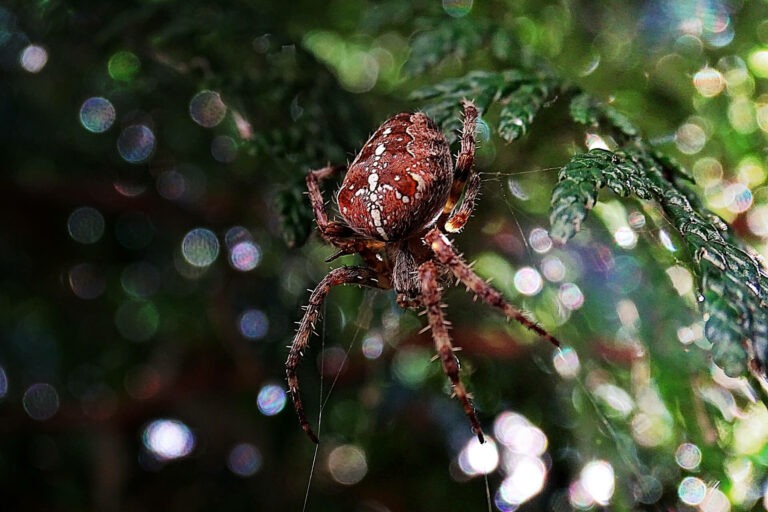
Coelho F. C., Amaya C. C. (2000): Measuring the heart rate of the spider, Aphonopelma hentzi: a non‐invasive technique. Physiological Entomology 25: 167-171.
FULL TEXT
Abstract
In this work we describe a non-invasive and precise technique to record the heartbeats of a spider. A linear output Hall effect transducer in conjunction with a small magnet was used to monitor the micromovements on the dorsal surface of the abdomen of the tarantula Aphonopelma hentzi (Girard) (Theraphosidae). The exoskeleton in this region is in direct contact with suspensory ligaments connected to the heart, and the dorsal cuticle of the opisthosoma moves with each heartbeat. The technique allowed the discrimination of the different stages of the spider’s cardiac cycle. The method can be also adapted for a smaller spider or other arthropods. We believe that the method proposed in this paper allows investigators to gain insights into a spider’s natural heart rate by gathering unbiased data with a non-invasive and very precise technique. We have found the resting heart rate of A. hentzi to be 5.6 ± 1.47 beats/min, which is lower than previously reported values.
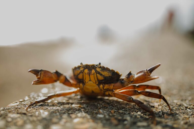
CRUSTACEANS
Wittwer C., Stoll S., Strand D., Vrålstad T., Nowak C., Thines M. (2018): eDNA-based crayfish plague monitoring is superior to conventional trap-based assessments in year-round detection probability. Hydrobiologia 807: 87-97.
FULL TEXT
Abstract
The crayfish plague disease agent Aphanomyces astaci is a major threat to European crayfish populations, leading to mass extinctions when spores are transmitted into habitats of native species by infected invasive crayfish species. Current methods for detecting crayfish plague in carrier crayfish populations depend on time-consuming capture of individuals and screening via molecular methods. Highly sensitive environmental DNA (eDNA) methods are a promising tool for rapid and cost-efficient monitoring of pathogens in freshwater systems directly in water samples. For evaluating the usefulness of eDNA for A. astaci detection, the trap-based crayfish plague monitoring followed by qPCR screening of tissue samples was compared to an eDNA-based system to detect A. astaci-spores at a stream inhabited by an infected carrier crayfish population of Pacifastacus leniusculus. The presence of A. astaci was confirmed at all investigated sites with both sample types. Both methods showed comparable A. astaci prevalence, with the eDNA method being applicable across a longer annual time span, including winter, with greater reliability than the conventional method. Given the speed and reliability of the eDNA method for crayfish plague detection, this method might be the best choice for routine monitoring and evaluation of crayfish habitats to hinder the disease spread.
Sieber N., Hartikainen H., Krieg R., Zenker A., Vorburger C. (2021): Parasite DNA detection in water samples enhances crayfish plague monitoring in asymptomatic invasive populations. Biological Invasions 24: 281-297.
FULL TEXT
Abstract
Invasive species can facilitate the spread of pathogens by first providing asymptomatic host reservoirs, and then driving disease outbreaks in native populations through pathogen spillover. An example of this are invasive crayfish species in Europe (Faxonius limosus, Pacifastacus leniusculus, Procambarus clarkii), which carry the deadly plague agent (Aphanomyces astaci). Effective disease management requires comprehensive monitoring, however, pathogen detection in carrier populations with low pathogen prevalence and intensities is challenging. We simultaneously collected and analysed crayfish tissue samples of invasive crayfish populations and water samples to compare A. astaci detection in different sample types using quantitative PCR. Combined, the two sampling methods revealed A. astaci presence with DNA concentrations above limit of detection (LOD; the lowest concentration which can be detected with reasonable certainty) in 13 of 23 invasive crayfish populations. In four additional sites, A. astaci DNA concentrations below LOD were found in water. In four populations only were A. astaci concentrations above LOD detected in both sample types and in three populations in concentrations above LOD in tissue but below LOD in water. The likely reason for these discrepancies is the low A. astaci prevalence and concentration in resistant invasive crayfish, which limit detection reliability. Consistency may be improved by timing surveys with seasonal periods of high A. astaci abundance and by increasing water sampling effort. Considering the ease of collecting eDNA samples, compared to crayfish tissue sampling, eDNA methods would facilitate frequent and comprehensive surveys. However, remaining uncertainties in eDNA-based detection reveal the relevance of combining monitoring tools to improve detection of invasive pathogens and their management.
Basso A., Paolini V., Ghia D., Fea G., Toson M., Pretto T. (2023): Cuticular swabs and eDNA as non-invasive sampling techniques to monitor Aphanomyces astaci in endangered white-clawed crayfish (Austropotamobius pallipes complex). Diversity 15: 279.
FULL TEXT
Abstract
In endangered crayfish conservation projects, it is paramount to map the distribution of the causative agent of crayfish plague, Aphanomyces astaci, in native populations. Considering the inapplicability of the destructive cuticular sampling protocol for monitoring endangered populations, we explored the use of non-invasive sampling techniques to detect this pathogen with molecular assays. In the present study, we exploited environmental DNA (testing increasing water volumes combined with different filter porosities) and cuticular swabs to collect A. astaci DNA. In addition, we evaluated the impact of the storage method on DNA preservation during field activities. After the first evaluations performed on both highly infected Austropotamobius pallipes and carrier Procambarus clarkii specimens in laboratory conditions, these sampling techniques were applied to wild populations of white-clawed crayfish. Our findings highlight better results with the filtration of 5 L of water with filters of 2.7 µm porosity for eDNA analysis and demonstrate that cuticular swabbing is equally effective as the World Organisation of Animal Health’s protocol. Storage in absolute ethanol proved to be the best solution to preserve swabs and filter samples for up to a week at room temperature. In conclusion, we suggest an integration of both sampling methods when monitoring A. astaci for conservation purposes.
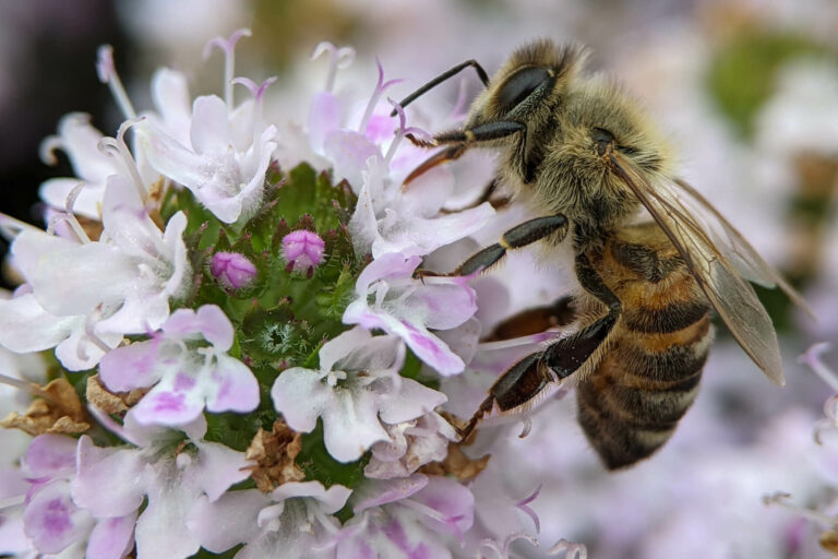
INSECTS
Schilling F., Dworschak K., Schopf R., Kühn R., Glaser S. J., Haase A. (2012): Non-invasive lipid measurement in living insects using NMR microscopy. Journal of Experimental Biology 215: 3137-3141.
FULL TEXT
Abstract
Nuclear magnetic resonance (NMR) microscopy allows us to image and quantify the distribution of NMR-active nuclei in living specimens. Using high-field NMR microscopy at a magnetic field strength of 14.1 T and strong gradients up to 3 T m−1, we show that separation of fat and water nuclear resonances in living insects can be achieved. In contrast to destructive conventional photometric and mass measurements, we demonstrate exemplarily in the European spruce bark beetle that NMR can be efficiently used to quantify absolute fat and water content in living insects. Additionally, anatomic images with a spatial in-plane resolution up to 10 μm and with high soft tissue contrast were acquired. We demonstrate that fat distribution and fat consumption of living insects can be obtained by magnetic resonance imaging (MRI). This enables future research to address questions where single individuals have to be measured several times, which is not possible with conventional destructive methods.
Goulson D., O’Connor S., Park K. J. (2018): The impacts of predators and parasites on wild bumblebee colonies. Ecological Entomology 43: 168-181.
FULL TEXT
Abstract
The study of wild bumblebee nests has been hindered by the difficulty in locating and observing them. Here, 47 wild nests were located using a sniffer dog and volunteers. The entrances to 32 nests were filmed continuously to identify successful nests (those that produced gynes) and observe vertebrate species interactions. Of the 47 nests, 71% and 21% produced gynes in 2010 and 2011, respectively. A total of 39 vertebrate species were filmed at entrances but the majority did not interact with the nests. Great tits (Parus major) depredated or attempted to depredate bees on 32 occasions at the entrances to 10 nests, something that has not previously been described. Small mammals were very often recorded accessing entrances to bumblebee nests, but whether they depredated bees was not known, and frequently visited nests were no less likely to produce gynes. Eight nests were entered by adult wax moths, Aphomia sociella. The faeces of 1179 workers from 29 Bombus terrestris nests were screened microscopically for parasites. Crithidia bombi infections were apparent in 49% of worker bees, while Nosema bombi and Apicystis bombi were present in 5.5% and 0.68% of bees, respectively. Nests with a high prevalence of C. bombi infection were less likely to produce gynes, the first evidence of a direct impact of this common parasite on bumblebee colony reproduction in wild nests. Overall, our data indicate that bumblebee nests are at the heart of a rich web of interactions between many different predator and parasite species.
Davis A. K. (2020): Evaluating cardiac reactions of monarch butterflies to human handling across three life stages. The Journal of the Lepidopterists’ Society 74: 43-50.
FULL TEXT
Abstract
Humans have an especially close connection with the monarch butterfly, Danaus plexippus, and this connection often involves direct handling of larvae, pupae or adults. Monarchs are frequently handled in citizen science programs, education and outreach activities, classroom demonstrations, and when rearing larvae in captivity. A pertinent question is whether monarchs perceive this human handling in the same way as being attacked by a predator, i.e. does it trigger a physiological fight-or-flight reaction? Here, I investigated if brief periods of handling monarch larvae, pupae, or adults elevates their heart rate (a central component of the insect fight-or-flight response), using non-destructive methods: either microscopy or an electronic heart monitor designed for invertebrates. Results are presented from separate investigations on each life stage. Three minutes of gentle handling caused a significant elevation (20%) in heart rates of 30 fifth-instar larvae. Mild physical disturbance for 20 seconds to a collection of 57 pupae elicited brief bursts of heart contractions (∼88 beats/min), a rate which is three times higher than typical contraction periods at this stage. Adult monarchs captured during fall migration (n=72) showed minor heart rate elevations (6%) after 3 minutes of gentle handling for tagging purposes. The differences in reactions across life stages found here is helpful for elucidating the relative sensitivity of each stage to handling. More importantly, these results elucidate the unseen physiological impact of direct human contact on developing monarchs (larvae and pupae); elevation in heart rate is an integral part of the fight-or-flight reaction in animals and a universal sign that an animal perceives an imminent threat. In this sense then, developing monarchs appear to perceive human handling as a threat, which may alter our (human) perception of this practice.
Davis A. K., Clancy K. M., Sasaki T. (2021): How to take an ant’s pulse: a procedure for non‐destructively monitoring baseline and stimulated heart rate in Formicidae. Entomologia Experimentalis et Applicata 169: 807-812.
FULL TEXT
Abstract
Ants, like other insects, have a heart that pumps hemolymph rhythmically. We designed an apparatus and procedure to non-destructively monitor the rate of cardiac contractions in ants, using a modified light microscope and infrared light. This allowed us to obtain the first baseline heart rate data on three ant species, collected in Georgia, USA. We next describe how ant heart rates can be stimulated, for comparing cardiac reactions among colonies, or for assessing stress responses. The procedures described here may be useful for other researchers interested in ant physiology.
Ribani A., Taurisano V., Utzeri V. J., Fontanesi L. (2022): Honey environmental DNA can be used to detect and monitor honey bee pests: Development of methods useful to identify Aethina tumida and Galleria mellonella infestations. Veterinary Sciences 9: 213.
FULL TEXT
Abstract
Environmental DNA (eDNA) contained in honey derives from the organisms that directly and indirectly have been involved in the production process of this matrix and that have played a role in the hive ecosystems where the honey has been produced. In this study we set up PCR-based assays to detect the presence of DNA traces left in the honey by two damaging honey bee pests: the small hive beetle (Aethina tumida) and the greater wax moth (Galleria mellonella). DNA was extracted from 82 honey samples produced in Italy and amplified using two specific primer pairs that target the mitochondrial gene cytochrome oxidase I (COI) of A. tumida and two specific primer pairs that target the same gene in G. mellonella. The limit of detection was tested using sequential dilutions of the pest DNA. Only one honey sample produced in Calabria was positive for A. tumida whereas about 66% of all samples were positively amplified for G. mellonella. The use of honey eDNA could be important to establish early and effective measures to contain at the local (e.g., apiary) or regional scales these two damaging pests and, particularly for the small hive beetle, to prevent its widespread diffusion.
Doherty J. F., Bhattarai U. R., Ferreira S., Poulin R., Gemmell N. J., Dowle E. J. (2023): The proof is in the poo: Non‐invasive method to detect endoparasitic infection. Molecular Ecology Resources.
FULL TEXT
Abstract
Almost every animal trait is strongly associated with parasitic infection or the potential exposure to parasites. Despite this importance, one of the greatest challenges that researchers still face is to accurately determine the status and severity of the endoparasitic infection without killing and dissecting the host. Thus, the precise detection of infection with minimal handling of the individual will improve experimental designs in live animal research. Here, we quantified extracellular DNA from two species of endoparasitic worm that grow within the host body cavity, hairworms (phylum Nematomorpha) and mermithids (phylum Nematoda), from the frass of their insect host, a cave wētā (Orthoptera: Rhaphidophoridae) and an earwig (Dermaptera: Forficulidae), respectively. Frass collection was done at two successive time periods, to test if parasitic growth correlated with relative DNA quantity in the frass. We developed and optimized two highly specific TaqMan assays, one for each parasite-specific DNA amplification. We were able to detect infection prevalence with 100% accuracy in individuals identified as infected through post-study dissections. An additional infection in earwigs was detected with the TaqMan assay alone, probably because some worms were either too small or degraded to observe during dissection. No difference in DNA quantity was detected between sampling periods, although future protocols could be refined to support such a trend. This study demonstrates that a noninvasive and minimally stressful method can be used to detect endoparasitic infection with greater accuracy than dissection alone, helping improve protocols for live animal studies.