MORPHOLOGY: AMPHIBIANS
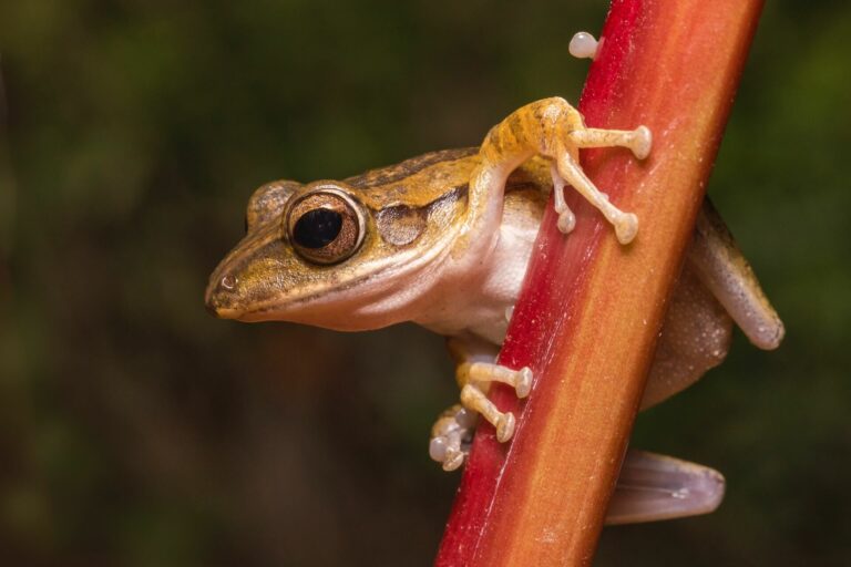
FROGS AND TOADS
Borzée A., Park S., Kim A., Kim H. T., Jang Y. (2013): Morphometrics of two sympatric species of tree frogs in Korea: a morphological key for the critically endangered Hyla suweonensis in relation to H. japonica. Animal Cells and Systems 17: 348-356.
FULL TEXT
Abstract
Although DNA taxonomy is readily available, morphological keys are still valuable for quick and easy identification of species on site. In Korea, Hyla japonica is widespread throughout the country, whereas Hyla suweonensis occurs in the lowlands of western central Korea. H. suweonensis is rapidly disappearing and was consequently designated as critically endangered by the Korean government. We measured 19 characters for male individuals of the two tree frog species to develop a morphological key for identification. Our morphometric analyses indicated that the two tree frog species differed significantly in means of all morphological characters. In general, H. suweonensis was smaller and more slender than H. japonica. Moreover, the distributions of five characters related to head width and the angle between eyes and ipsilateral nostrils did not overlap in the two species and may be used for species identification. Because the character differences between the two species are small, all five characters should be used together to reliably distinguish the two tree frog species. Besides being used as a morphological key, our result in size difference leads to several research questions about microhabitat niche selection and competition between the two Korean tree frog species.
Kim E., Sung H., Lee D., Kim G., Nam D., Kim E. (2017): Nondestructive skeletal imaging of Hyla suweonensis using micro-computed tomography. Asian Herpetological Research 8: 235-43.
FULL TEXT
Abstract
We successfully obtained 3D skeletal images of Hyla suweonensis, employing a nondestructive method by applying appropriate anesthesia and limiting the radiation dose. H. suweonensis is a tree frog endemic to Korea and is on the list of endangered species. Previous studies have employed caliper-based measurements and two-dimensional (2D) X-ray imaging for anatomical analyses of the skeletal system or bone types of H. suweonensis. In this work we reconstructed three-dimensional (3D) skeletal images of H. suweonensis, utilizing a nondestructive micro-computed tomography (micro-CT) with a short scan and low radiation dose (i.e. 4 min and 0.16 Gy). Importantly, our approach can be applied to the imaging of 3D skeletal systems of other endangered frog species, allowing both versatile and high contrast images of anatomical structures without causing any significant damages to the living animal.
Abdul Aziz M. F., Mohd M., Shohaimi S., Ab Ghani N. I., Fletcher C. (2021): Morphometric study of Kalophrynus palmatissimus at two forest reserves in Malaysia. Ecology and Evolution 11: 10741-10753.
FULL TEXT
Abstract
A research study on morphometrics of Kalophrynus palmatissimus (commonly known as Lowland Grainy Frog) at Ayer Hitam Forest Reserve (AHFR), Selangor and Pasoh Forest Reserve (PFR), Negeri Sembilan was carried out from 12 November 2016 to 13 September 2017. The study was to examine data on the morphometric traits of K. palmatissimus at the two forest reserves. 15 morphometric traits of K. palmatissimus that were taken by using vernier calipers. Frog surveys were done by using 15 and 18 nocturnal 400 m transect lines with an interval distance of 20 m at AHFR and PFR, respectively. The GPS coordinates for all frog samples were recorded to ensure the precise geographic location. In addition, five climatic data were recorded. The results showed that most morphometric traits in AHFR (n = 34) and PFR (n = 31) were positively correlated with each other. On the other hand, climatic factor, which was soil pH, had a significant positive influence on most of the morphometric traits (p < .01), except for tympanum diameter and upper eyelid width (p ≥ .05). Meanwhile, the temperature had a significantly negative influence on all morphometric traits (p < .01). General linear model (GLM) analysis showed that snout-vent length (SVL) influenced most morphometric traits (F ≤ 80.86, p < .01), except for hand length (HAL: F = 0.299, p > .05). Later, it was found that the snout-vent length of K. palmatissimus at AHFR was slightly larger than at PFR (AHFR: μ = 37.00 mm, SE = 1.16 c.f. PFR: μ = 30.29 mm, SE = 1.07). It showed that there were variations in morphometric traits of K. palmatissimus at AHFR and PFR. From PCA analysis, morphometric traits are grouped into two components for AHFR and PFR, respectively. In AHFR, head length, eye diameter, head width, internarial distance, interorbital distance, forearm length, tibia length, foot length, and thigh length were strongly correlated, while snout length and eye-nostril distance were strongly correlated. In PFR, eye diameter, head width, internarial distance, interorbital distance, foot length, and thigh length were strongly correlated, though snout length and eye-nostril distance were strongly correlated, hence, suggested that all morphometric traits grow simultaneously in K. palmatissimus with eye-nostril distance (EN), and snout length (SL) growing almost simultaneously at AHFR (r = .91) and PFR (r = .97). There is still a lack of available information regarding the distribution and morphometric studies of K. palmatissimus in Malaysia, especially at AHFR and PFR. This study showed 15 different morphometric traits of K. palmatisssimus between AHFR and PFR, with K. palmatissimus at AHFR were found to be slightly larger than at PFR.
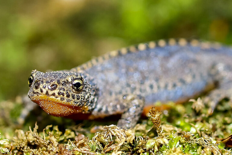
SALAMANDERS AND NEWTS
Alarcón‐Ríos L., Velo‐Antón G., Kaliontzopoulou A. (2017): A non‐invasive geometric morphometrics method for exploring variation in dorsal head shape in urodeles: sexual dimorphism and geographic variation in Salamandra salamandra. Journal of Morphology 278: 475-485.
FULL TEXT
Abstract
The study of morphological variation among and within taxa can shed light on the evolution of phenotypic diversification. In the case of urodeles, the dorso-ventral view of the head captures most of the ontogenetic and evolutionary variation of the entire head, which is a structure with a high potential for being a target of selection due to its relevance in ecological and social functions. Here, we describe a non-invasive procedure of geometric morphometrics for exploring morphological variation in the external dorso-ventral view of urodeles’ head. To explore the accuracy of the method and its potential for describing morphological patterns we applied it to two populations of Salamandra salamandra gallaica from NW Iberia. Using landmark-based geometric morphometrics, we detected differences in head shape between populations and sexes, and an allometric relationship between shape and size. We also determined that not all differences in head shape are due to size variation, suggesting intrinsic shape differences across sexes and populations. These morphological patterns had not been previously explored in S. salamandra, despite the high levels of intraspecific diversity within this species. The methodological procedure presented here allows to detect shape variation at a very fine scale, and solves the drawbacks of using cranial samples, thus increasing the possibilities of using collection specimens and alive animals for exploring dorsal head shape variation and its evolutionary and ecological implications in urodeles.
Arismendi I., Bury G., Zatkos L., Snyder J., Lindley D. (2021): A method to evaluate body length of live aquatic vertebrates using digital images. Ecology and Evolution 11: 5497-5502.
FULL TEXT
Abstract
Traditional methods to measure body lengths of aquatic vertebrates rely on anesthetics, and extended handling times. These procedures can increase stress, potentially affecting the animal’s welfare after its release. We developed a simple procedure using digital images to estimate body lengths of coastal cutthroat trout (Oncorhynchus clarkii clarkii) and larval coastal giant salamander (Dicamptodon tenebrosus). Images were postprocessed using ImageJ2. We measured more than 900 individuals of these two species from 200 pool habitats along 9.6 river kilometers. The percent error (mean ± SE) of our approach compared to the use of a traditional graded measuring board was relatively small for all length metrics of the two species. Total length of trout was −2.2% ± 1.0. Snout–vent length and total length of larval salamanders was 3.5% ± 3.3 and −0.6% ± 1.7, respectively. We cross-validated our results by two independent observers that followed our protocol to measure the same animals and found no significant differences (p > .7) in body size distributions for all length metrics of the two species. Our procedure provides reliable information of body size reducing stress and handling time in the field. The method is transferable across taxa and the inclusion of multiple animals per image increases sampling efficiency with stored images that can be reviewed multiple times. This practical tool can improve data collection of animal size over large sampling efforts and broad spatiotemporal contexts.


