MORPHOLOGY: ARTHROPODS
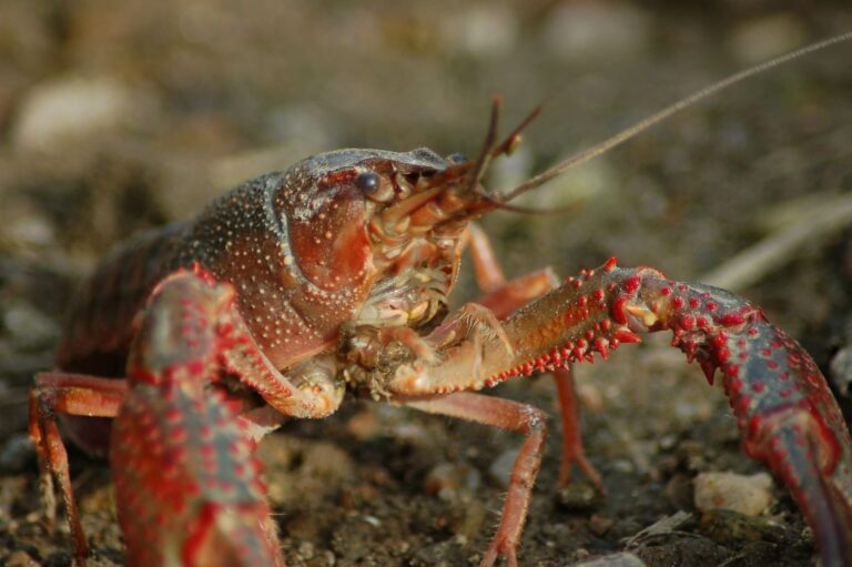
CRUSTACEANS
Ulikowski D., Krzywosz T. (2009): In vivo calibration of juvenile crayfish body length and weight with a photographic-computer method. Fisheries & Aquatic Life 17: 89-93.
FULL TEXT
Abstract
The aim of this work was to develop a photographic-computer method to determine in vivo the body length and weight of juvenile crayfish based on standards that describe the dependence curves of these characters. The measurement procedure that was developed requires one digital group photograph of a sample generally comprised of thirty or so individuals and an image scale, which is visible within the frame. A special program is used to analyze the image according to the scale on the image. Further, only the cephalothorax length (CL) of specimens from the studied sample that appear in the photograph is measured. Total length (TL) and weight (W) are then calculated with correlation formulae that were developed for the characters analyzed. The following dependencies were calculated from the formulae developed for two-month-old narrow-clawed crayfish, Astacus leptodactylus (Esch.) (n = 101, W = 225.9 ± 67.2 mg, TL = 18.7 ± 2.1 mm): TL = 1.739 CL + 1.1903, W = 0.1306 TL 2.533, W = 0.7635 CL 2.4476. All of the dependencies were highly significant statistically (P < 0.001).
Xue J., Liu H., Jiang T., Chen X., Yang J. (2022): Shape variation in the carapace of Chinese mitten crabs (Eriocheir sinensis H. Milne Edwards, 1853) in Yangcheng lake during the year-long culture period. The European Zoological Journal 89: 217-228.
FULL TEXT
Abstract
To evaluate whether ontogenetic development over the grow-out culture period can cause geographical plasticity of carapace shape in a certain region, geometric morphometric analysis was used, in this study, to determine the ontogenetic changes in the carapace morphology of Chinese mitten crabs (Eriocheir sinensis) originating from Yangcheng Lake, China, during a year of growth from coin-sized juveniles to market-sized adults. The morphological differences in the carapace throughout the year of culture were characterized using a 35-landmark point system. We used principal component analysis and linear discriminant analysis to determine morphological variation using thin-plate spline analysis and mesh deformation. During the growth process, the changes in the carapace were mainly concentrated in the third and fourth anterolateral teeth and on the M-shaped pattern. During the growth of male and female crabs throughout the year, the shape of the carapace changed considerably over the first six months. Afterwards, the shape of the carapace began to stabilize and could not be differentiated through discriminant analysis. This study is the first to use geometric morphometrics to analyze the ontogenetic changes in the carapace shape of E. sinensis crabs native to Yangcheng Lake. The results demonstrate that it takes a long time for the carapace morphology of native crabs to stabilize.
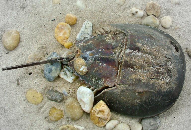
HORSESHOE CRABS
Yuen A. H. L., Kwok D. H. C., Kim S. W. (2019): Magnetic resonance imaging of the live tri-spine horseshoe crab (Tachypleus tridentatus). Arthropoda Selecta 28: 247-251.
FULL TEXT
Abstract
Tri-spine horseshoe crab (Tachypleus tridentatus) is one of the most extensively studied arthropods from both biological and paleontological perspectives due to its unique suite of anatomical features and as a useful modern analogue for fossil arthropod groups. To assist the study and documentation of this iconic taxon, thorough understanding of their anatomy is necessary. Traditional dissection approach to study the anatomy of tri-spine horseshoe crab is technically demanding and time-consuming, and causes loss of specimen integrity. Magnetic resonance imaging (MRI) have currently become more readily available for zoomorphological investigation. A growing body of digitally stored anatomical data has become available to assist with biological, morphological and pathological investigation, without destroying specimens. The objective of the present study is to provide an overview of the normal cross-sectional anatomy of the live tri-spine horseshoe crab using T1W and T2W MRI, along with dissection images. MRI scan of 4 living tri-spine horseshoe crabs were performed by 1.5T MRI scanner. The resulting images provided excellent detail of major anatomical structures of live tri-spine horseshoe crabs. The illustrations in the present study provides an initial reference to evaluate anatomical structures of the tri-spine horseshoe crab on MR images.
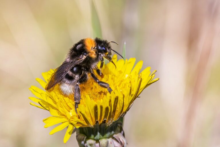
INSECTS
Koehler C., Liang Z., Gaston Z., Wan H., Dong H. (2012): 3D reconstruction and analysis of wing deformation in free-flying dragonflies. Journal of Experimental Biology 215: 3018-3027.
FULL TEXT
Abstract
Insect wings demonstrate elaborate three-dimensional deformations and kinematics. These deformations are key to understanding many aspects of insect flight including aerodynamics, structural dynamics and control. In this paper, we propose a template-based subdivision surface reconstruction method that is capable of reconstructing the wing deformations and kinematics of free-flying insects based on the output of a high-speed camera system. The reconstruction method makes no rigid wing assumptions and allows for an arbitrary arrangement of marker points on the interior and edges of each wing. The resulting wing surfaces are projected back into image space and compared with expert segmentations to validate reconstruction accuracy. A least squares plane is then proposed as a universal reference to aid in making repeatable measurements of the reconstructed wing deformations. Using an Eastern pondhawk (Erythimus simplicicollis) dragonfly for demonstration, we quantify and visualize the wing twist and camber in both the chord-wise and span-wise directions, and discuss the implications of the results. In particular, a detailed analysis of the subtle deformation in the dragonfly’s right hindwing suggests that the muscles near the wing root could be used to induce chord-wise camber in the portion of the wing nearest the specimen’s body. We conclude by proposing a novel technique for modeling wing corrugation in the reconstructed flapping wings. In this method, displacement mapping is used to combine wing surface details measured from static wings with the reconstructed flapping wings, while not requiring any additional information be tracked in the high speed camera output.
Bourne D. R., Kyle C. J., LeBlanc H. N., Beresford D. (2019): A rapid, non-invasive method for measuring live or preserved insect specimens using digital image analysis. Forensic Science International: Synergy 1: 140-145.
FULL TEXT
Abstract
The measurement of insects is an important component of many entomological applications, including forensic evidence, where larvae size is used as a proxy for developmental stage, and hence time since colonization/death. Current methods for measuring insects are confounded by varying preservation techniques, biased and non-standardized measurements, and often a lack of sample size given practical constraints. Towards enhanced accuracy and precision in measuring live insects to help avoid these variables, and that allows for different measurements to be analyzed, we developed a non-invasive, digital method using widely available free analytical software to measure live blow fly larvae. Using crime scene photographic equipment currently standard in investigation protocols, we measured the live length of 282 Phormia regina larvae. Repeated measurements of maggots, for all instars, were performed for several orientations and images. Most accurate measurements were obtained when maggots were oriented in their natural full extension. Killed specimens resulted in greater length measurements (Mean 1.79 ± 1.11 mm) when compared to live length. Herein, we report a technically simple, fast, and accurate measurement technique adapted for field and lab-based measurements, as well as, a simple linear equation for conversion of live length to standard killed length measurements. We propose this method be utilized for the standardization of forensic entomological evidence collection and development model creation.
Mungee M., Athreya R. (2020): Rapid photogrammetry of morphological traits of free‐ranging moths. Ecological Entomology 45: 911-923.
FULL TEXT
Abstract
Photogrammetric studies of free-ranging animals are limited to mammals and birds. Recent advances in insect photogrammetry, including 3D imaging, are entirely associated with museum specimens. We present a rapid, simple, accurate, and inexpensive morphometric method targeting thousands of free-ranging insects attracted to light screens using images taken without collecting a specimen or even constraining the individual in any manner. A reference grid printed on the screen is used to calibrate the images for shape and size without prior knowledge of the camera-subject configuration. The method requires only inexpensive, off-the-shelf, consumer equipment, and freely available programming (R statistical language) and image processing (ImageMagick) tools. We demonstrate the efficacy of the method using a dataset of 3675 images of free-ranging hawkmoths (Lepidoptera: Sphingidae) imaged in natural repose on a screen. We show that this method introduces no bias and has a high degree of correspondence with traditional morphometry using collected specimens. We also propose error metrics, which quantify the calibration quality and identify images with poor data. Although this method is particularly suited for the hyperdiverse moth community, which dominates the dynamics of many terrestrial ecosystems, it can be used for other phototropic taxa identifiable on an image to (morpho)-species. It will help in accumulating reliable trait data from hundreds of thousands of individual insects without any expenditure on specimen collection. It is particularly suited for studies which require multi-epoch, multi-locate sampling like investigations into ecosystem stability, climate change, and community assembly.
Lertvilai P., Jaffe J. S. (2022): In situ size and motility measurement of aquatic invertebrates with an underwater stereoscopic camera system using tilted lenses. Methods in Ecology and Evolution 13: 1192-1200.
FULL TEXT
Abstract
In situ observation of traits of aquatic organisms, including size and motility, requires three-dimensional measurements that are commonly done with a stereoscopic imaging system. However, to observe traits of small aquatic invertebrates, the imaging system requires relatively high magnification, which results in a small overlapping volume between the two cameras of a conventional stereoscopic system. The provision of a larger shared volume would therefore be of great advantage, especially, when the organism abundance is low. We implement a stereoscopic system that utilizes a tilted lens approach, known as the Scheimpflug principle, to increase the common imaging volume of two cameras. The system was calibrated and tested in the laboratory and then deployed in a saltmarsh to observe water boatmen Trichocorixa californica. Processing of the image data from the field deployments resulted in the simultaneous estimation of the traits of body length and swimming speed of the aquatic insects. Our stereo setup with tilted lenses increased the sampling volume by 3.1 times compared to a traditional stereo setup with the same optical parameters. The in situ data and subsequent processing reveal that the instrument can capture stereoscopic images that resolve both body length and swimming speed of the aquatic insects. Results indicate that the relationship between the body length and the swimming speed of the water boatmen is linear in the log–log space with an exponent of 0.81±0.12. Furthermore, the insects experience Reynold’s number in the range of 100-103. Our results demonstrated that the system can be used to observe key traits of small aquatic organisms in an ecologically relevant context. This work expands the capability of underwater imaging systems to measure important traits of an individual aquatic invertebrate in its natural environment and aids in providing a trait-based approach to zooplankton ecology.


