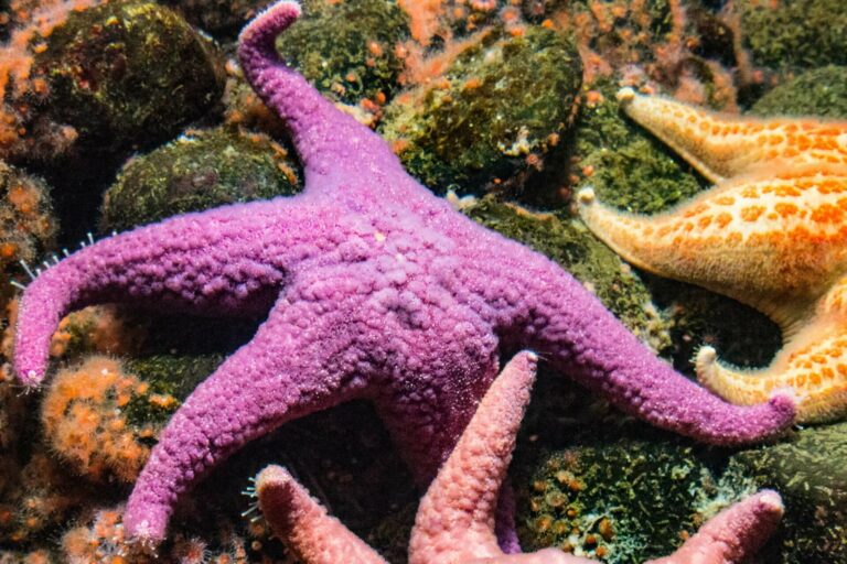
MORPHOLOGY: ECHINODERMS
STARFISH
Sigl R., Imhof H., Settles M., Laforsch C. (2013): A novel, non-invasive and in vivo approach to determine morphometric data in starfish. Journal of Experimental Marine Biology and Ecology 449: 1-9.
FULL TEXT
Abstract
Starfish (Echinodermata: Asteroidea) are present in most benthic ocean habitats and play an important ecological role as keystone species or by dominating through sheer individual numbers. In order to assess nutritional and reproductive states in ecological studies on asteroids, invasive techniques to calculate organ indices are conventionally used. We present a non-invasive method that enables imaging and morphometric measurements in starfish in vivo. We used a clinical 1.5 T magnetic resonance imaging (MRI) scanner to produce sectional images of three starfish species and employed these image stacks to generate 3D models of the pyloric ceca, gonads and the endoskeleton. In comparison to pre-clinical MRI scanners, that provide higher resolutions, clinical MRI is not limited to small objects, but allows the investigation of larger samples such as the starfish used in the present study. Volume data from MRI-based 3D reconstructions were compared to conventional invasive measurement techniques as well as high resolution MRI scans and were tested for inter-observer effects. Here we show that MRI is a suitable method for precise imaging and volumetric measurements in fixed and living marine specimens. Compared to other methods, it allows not only the production of time series data on single individuals as well as populations, but also non-destructive analyses of valuable specimens, such as museum material.


