MORPHOLOGY: MAMMALS
BATS
Schmieder D. A., Benítez H. A., Borissov I. M., Fruciano C. (2015): Bat species comparisons based on external morphology: a test of traditional versus geometric morphometric approaches. Plos One 10: e0127043.
FULL TEXT
Abstract
External morphology is commonly used to identify bats as well as to investigate flight and foraging behavior, typically relying on simple length and area measures or ratios. However, geometric morphometrics is increasingly used in the biological sciences to analyse variation in shape and discriminate among species and populations. Here we compare the ability of traditional versus geometric morphometric methods in discriminating between closely related bat species – in this case European horseshoe bats (Rhinolophidae, Chiroptera) – based on morphology of the wing, body and tail. In addition to comparing morphometric methods, we used geometric morphometrics to detect interspecies differences as shape changes. Geometric morphometrics yielded improved species discrimination relative to traditional methods. The predicted shape for the variation along the between group principal components revealed that the largest differences between species lay in the extent to which the wing reaches in the direction of the head. This strong trend in interspecific shape variation is associated with size, which we interpret as an evolutionary allometry pattern.
CARNIVORES
de Bruyn P. N., Bester M. N., Carlini A. R., Oosthuizen W. C. (2009): How to weigh an elephant seal with one finger: a simple three-dimensional photogrammetric application. Aquatic Biology 5: 31-39.
FULL TEXT
Abstract
Several studies have developed photogrammetric techniques for indirect mass estimation of seals. Unfortunately, these techniques are often narrowly delineated for specific field scenarios or species. Many require sophisticated, custom-designed equipment or analytical tools, limiting their applicability. We aimed to devise a photogrammetric technique for accurate volume/mass estimation of seals under a variety of field scenarios without manipulation of the animal and with minimal equipment. We used Photomodeler Pro 3-dimensional modelling software to estimate the mass of 53 weighed southern elephant seals Mirounga leonina. The method is centred on animal volume estimation in relation to the 3-dimensional area around it, rather than features of the animal itself, an approach that liberates limitations associated with earlier studies. No morphometric body measures are required for such volume/mass estimation. We offer predictive equations that allow high confidence in mass estimates relative to measured mass (95% confidence interval of mean deviation from measured mass is from ±1.34 to ±3.83% depending on the field scenario). A single photographer with a measuring stick and non-customised digital photographic equipment can use this technique to determine the mass of an elephant seal anywhere in the field with the push of a button.
Meise K., Mueller B., Zein B., Trillmich F. (2014): Applicability of single-camera photogrammetry to determine body dimensions of pinnipeds: Galapagos sea lions as an example. Plos One 9: e101197.
FULL TEXT
Abstract
Morphological features correlate with many life history traits and are therefore of high interest to behavioral and evolutionary biologists. Photogrammetry provides a useful tool to collect morphological data from species for which measurements are otherwise difficult to obtain. This method reduces disturbance and avoids capture stress. Using the Galapagos sea lion (Zalophus wollebaeki) as a model system, we tested the applicability of single-camera photogrammetry in combination with laser distance measurement to estimate morphological traits which may vary with an animal’s body position. We assessed whether linear morphological traits estimated by photogrammetry can be used to estimate body length and mass. We show that accurate estimates of body length (males: ±2.0%, females: ±2.6%) and reliable estimates of body mass are possible (males: ±6.8%, females: 14.5%). Furthermore, we developed correction factors that allow the use of animal photos that diverge somewhat from a flat-out position. The product of estimated body length and girth produced sufficiently reliable estimates of mass to categorize individuals into 10 kg-classes of body mass. Data of individuals repeatedly photographed within one season suggested relatively low measurement errors (body length: 2.9%, body mass: 8.1%). In order to develop accurate sex- and age-specific correction factors, a sufficient number of individuals from both sexes and from all desired age classes have to be captured for baseline measurements. Given proper validation, this method provides an excellent opportunity to collect morphological data for large numbers of individuals with minimal disturbance.
Krause D. J., Hinke J. T., Perryman W. L., Goebel M. E., LeRoi D. J. (2017): An accurate and adaptable photogrammetric approach for estimating the mass and body condition of pinnipeds using an unmanned aerial system. Plos One 12: e0187465.
FULL TEXT
Abstract
Measurements of body size and mass are fundamental to pinniped population management and research. Manual measurements tend to be accurate but are invasive and logistically challenging to obtain. Ground-based photogrammetric techniques are less invasive, but inherent limitations make them impractical for many field applications. The recent proliferation of unmanned aerial systems (UAS) in wildlife monitoring has provided a promising new platform for the photogrammetry of free-ranging pinnipeds. Leopard seals (Hydrurga leptonyx) are an apex predator in coastal Antarctica whose body condition could be a valuable indicator of ecosystem health. We aerially surveyed leopard seals of known body size and mass to test the precision and accuracy of photogrammetry from a small UAS. Flights were conducted in January and February of 2013 and 2014 and 50 photogrammetric samples were obtained from 15 unrestrained seals. UAS-derived measurements of standard length were accurate to within 2.01 ± 1.06%, and paired comparisons with ground measurements were statistically indistinguishable. An allometric linear mixed effects model predicted leopard seal mass within 19.40 kg (4.4% error for a 440 kg seal). Photogrammetric measurements from a single, vertical image obtained using UAS provide a noninvasive approach for estimating the mass and body condition of pinnipeds that may be widely applicable.
Galimberti F., Sanvito S., Vinesi M. C., Cardini A. (2019): “Nose‐metrics” of wild southern elephant seal (Mirounga leonina) males using image analysis and geometric morphometrics. Journal of Zoological Systematics and Evolutionary Research 57: 710-720.
FULL TEXT
Abstract
The elephant seal (genus Mirounga) proboscis is a textbook example of an exaggerated secondary sexual trait, whose function is debated. The proboscis can be related to sexual status advertising, emission of aggressive vocalizations, and/or female mating choice. The study of the proboscis is complicated, because it is a soft trait that needs to be studied when males vocalize and it is thus expanded. Here, we combined field stimulation experiments, 2D photogrammetry, and geometric morphometrics, to study the proboscis of wild elephant seals in natural conditions. The goal of our study was twofold: (a) to demonstrate that photogrammetry and geometric morphometrics can be effectively applied in the field to wild, large, non-sedated mammals; (b) to study the proboscis shape development during maturation. We found that it is possible to accurately estimate the proboscis size and shape using photographs of vocalizing males taken in the field. Moreover, we showed that mature and non-mature males differ not only in proboscis size but also in its shape, a difference that is largely allometric and can have important effects on the frequency structure and individual signature of male vocalizations. These results open new avenues for future research on this enigmatic structure, its function in aggressive displays and potential role in sexual selection, and also exemplify a very promising approach, that could be applied in field studies of other large mammals.
Sanvito S., Sanvito R., Galimberti F. (2019): Rapid noninvasive field estimation of body length of female elephant seals (Mirounga leonina). Marine Mammal Science 35: 345-352.
FULL TEXT
Excerpt
Here, we present a method to measure wild seals noninvasively that is implemented with inexpensive instruments, requires no postprocessing, and produces size estimates that can be used immediately. We calculated the error of the method by measuring objects of known size, we applied the method to female elephant seals and calculated repeatability of field measurements, and we estimated empirical equations that permit conversion among the different measurements that can be obtained. We discuss the advantages of this new method in comparison with photogrammetric approaches. Our measurement method was based on the application of simple trigonometry to measured distances and angles.
Tarugara A., Clegg B. W., Gandiwa E., Muposhi V. K., Wenham C. M. (2019): Measuring body dimensions of leopards (Panthera pardus) from camera trap photographs. PeerJ 7: e7630.
FULL TEXT
Abstract
Measurement of body dimensions of carnivores usually requires the chemical immobilization of subjects. This process can be dangerous, costly and potentially harmful to the target individuals. Development of an alternative, inexpensive, and non-invasive method therefore warrants attention. The objective of this study was to test whether it is possible to obtain accurate measurements of body dimensions of leopards from camera trap photographs. A total of 10 leopards (Panthera pardus) were captured and collared at Malilangwe Wildlife Reserve, Zimbabwe from May 7 to June 20, 2017 and four body measurements namely shoulder height, head-to-tail, body, and tail length were recorded. The same measurements were taken from 101 scaled photographs of the leopards recorded during a baited-camera trapping (BCT) survey conducted from July 1 to October 22, 2017 and differences from the actual measurements calculated. Generalized Linear Mixed Effects Models were used to determine the effect of type of body measurement, photographic scale, posture, and sex on the accuracy of the photograph-based measurements. Type of body measurement and posture had a significant influence on accuracy. Least squares means of absolute differences between actual and photographic measurements showed that body length in the level back-straight forelimb-parallel tail posture was measured most accurately from photographs (2.0 cm, 95% CI [1.5–2.7 cm]), while head-to-tail dimensions in the arched back-bent forelimb-parallel tail posture were least accurate (8.3 cm, 95% CI [6.1–11.2 cm]). Using the BCT design, we conclude that it is possible to collect accurate morphometric data of leopards from camera trap photographs. Repeat measurements over time can provide researchers with vital body size and growth rate information which may help improve the monitoring and management of species of conservation concern, such as leopards.
Alvarado D. C., Robinson P. W., Frasson N. C., Costa D. P., Beltran R. S. (2020): Calibration of aerial photogrammetry to estimate elephant seal mass. Marine Mammal Science 36: 1347-1355.
FULL TEXT
Excerpt
In summary, we determined the error associated with using UAS photogrammetry to estimate northern elephant seal mass. Photogrammetric measurements from a single, vertical image obtained using UAS provide a promising approach for estimating the body mass of pinnipeds and similar approaches can be used to find species-specific calibration equations.
Hodgson J. C., Holman D., Terauds A., Koh L. P., Goldsworthy S. D. (2020): Rapid condition monitoring of an endangered marine vertebrate using precise, non-invasive morphometrics. Biological Conservation 242: 108402.
FULL TEXT
Abstract
Understanding causes of population change is critical for conservation. Quantifying these causes can be difficult, especially for hard to sample animals like marine vertebrates (e.g. pinnipeds). One solution is to investigate spatiotemporal differences in a species’ body condition by measuring body size and mass. Collecting traditional morphological measurements is risky and labour intensive, making less invasive and more efficient techniques desirable. Using Australian sea lions (Neophoca cinerea) of known size and mass as a case study, we tested the suitability of using drone-derived photogrammetry to estimate morphological measurements and assess body condition. Drone-derived measurements were precise and without bias. Animal mass was highly correlated with the 2-dimensional and 3-dimensional measurements of simplified area and volume, explaining >77% and >84% (all P < .01) of the variation in mass, depending on the age-sex class. The juvenile class exhibited the strongest associations (both 2D and 3D R2 > 0.99). Using each measurement as a proxy for mass, we calculated body condition indices for each class by standardising the variables by animal length. Photogrammetric indices ranked individuals comparably to those generated from ground-collected data (rs = 0.77–1, depending on age-sex class). Our technique provides a workflow for the non-invasive collection of morphometric data to quantify animal condition, which is transferrable to other pinniped species with species-specific calibration. It will also facilitate the efficient collection of morphometric data of vertebrates from remotely sensed imagery.
Rasmussen G., Smultea M., Cloutier T., Giordano A., Kaplin B., Willey L. (2021): Quantitative photogrammetric methodology for measuring mammalian belly score in the painted dog. Plos One 16: e0261171.
FULL TEXT
Abstract
The use of “belly scoring” can offer a novel, non-invasive objective management tool to gauge food intake between individuals, groups, and populations, and thus, population fitness. As food availability is increasingly affected by predation, ecological competition, climate change, habitat modification, and other human activities, an accurate belly scoring tool can facilitate comparisons among wildlife populations, serving as an early warning indicator of threats to wildlife population health and potential population collapse. In social species, belly scores can also be a tool to understand social behavior and ranking. We developed and applied the first rigorous quantitative photogrammetric methodology to measure belly scores of wild painted dogs (Lycaon pictus). Our methodology involves: (1) Rigorous selection of photographs of the dorso/lateral profile of individuals at a right angle to the camera, (2) photogrammetrically measuring belly chord length and “belly drop” in pixels, (3) adjusting belly chord length as a departure from a standardized leg angle, and (4) converting pixel measurements to ratios to eliminate the need to introduce distance from the camera. To highlight a practical application, this belly score method was applied to 631 suitable photographs of 15 painted dog packs that included 186 individuals, all collected between 2004–2015 from allopatric painted dog populations in and around Hwange (n = 462) and Mana Pools National Parks (n = 169) in Zimbabwe. Variation in mean belly scores exhibited a cyclical pattern throughout the year, corresponding to biologically significant patterns to include denning demand and prey availability. Our results show significant differences between belly scores of the two different populations we assessed, thus highlighting food stress in the Hwange population. In the face of growing direct and indirect anthropogenic disturbances, this standardised methodology can provide a rapid, species-specific non-invasive management tool that can be applied across studies to rapidly detect emergent threats.
Eder E. B., Almonacid J. S., Delrieux C., Lewis M. N. (2022): Body volume and mass estimation of southern elephant seals using 3D range scanning and neural network models. Marine Mammal Science 38: 1037-1049.
FULL TEXT
Abstract
Direct measures of body mass of marine mammals are logistically complicated to obtain even for pinnipeds. An alternative method for mass estimation based on 3D imaging technology and automated processing algorithms, was tested in southern elephant seals (Mirounga leonina). Two models of artificial neural networks (ANN)—nonlinear neural network and self-organizing maps—were trained to compute the volume and to estimate the mass of the digital models, previously obtained by scanning individuals with an infrared light sensor. Body mass estimates were as accurate (mean % error = 4.4) as estimates in previous photogrammetry studies in southern elephant seals and the mass predictive ability of the trained ANN was higher (99% of the variance explained) than other predictive models using photogrammetry in pinniped studies. While this method has proven to produce accurate body mass estimates, it also overcame some of the constraints of other indirect techniques, avoiding animal disturbance caused by physical restraint or chemical immobilization, minimizing risks, and capitalizing on the time working in the field. The results of these estimations were promising, which shows that the proposed methodology can provide adequate results with lower logistic and computational requirements.
Carroll D., Infantes E., Pagan E. V., Harding K. C. (2024): Approaching a population‐level assessment of body size in pinnipeds using drones, an early warning of environmental degradation. Remote Sensing in Ecology and Conservation.
FULL TEXT
Abstract
Body mass is a fundamental indicator of animal health closely linked to survival and reproductive success. Systematic assessment of body mass for a large proportion of a population can allow early detection of changes likely to impact population growth, facilitating responsive management and a mechanistic understanding of ecological trends. One challenge with integrating body mass assessment into monitoring is sampling enough animals to detect trends and account for individual variation. Harbour seals (Phoca vitulina) are philopatric marine mammals responsive to regional environmental changes, resulting in their use as an indicator species. We present a novel method for the non-invasive and semi-automatic assessment of harbour seal body condition, using unoccupied aerial vehicles (UAVs/drones). Morphological parameters are automatically measured in georeferenced images and used to estimate volume, which is then translated to estimated mass. Remote observations of known individuals are utilized to calibrate the method. We achieve a high level of accuracy (mean absolute error of 4.5 kg or 10.5% for all seals and 3.2 kg or 12.7% for pups-of-the-year). We systematically apply the method to wild seals during the Spring pupping season and Autumn over 2 years, achieving a near-population-level assessment for pups on land (82.5% measured). With reference to previous mark-recapture work linking Autumn pup weights to survival, we estimate mean expected probability of over-winter survival (mean = 0.89, standard deviation = 0.08). This work marks a significant step forward for the non-invasive assessment of body condition in pinnipeds and could provide daily estimates of body mass for thousands of individuals. It can act as an early warning for deteriorating environmental conditions and be utilized as an integrative tool for wildlife monitoring. It also enables estimation of yearly variation in demographic rates which can be utilized in parameterizing models of population growth with relevance for conservation and evolutionary biology.
Stone T. C., Davis K. J. (2024): Using unmanned aerial vehicles to estimate body volume at scale for ecological monitoring. Methods in Ecology and Evolution.
FULL TEXT
Abstract
Demographic data are essential to construct mechanistic models to understand how populations change over time and in response to global threats like climate change. Existing demographic data are either lacking or insufficient for many species, particularly those that are challenging to obtain direct measurements from that can be used to estimate demographic rates, like marine mammals. A method for collecting accurate demographic data to construct robust demographic models at scale would fill this knowledge gap for difficult-to-access species. We introduce a novel, non-invasive method to estimate the 3D body size (volume) of pinnipeds (seals, sea lions and walruses) that will allow monitoring at high spatial and temporal scales. Our method integrates 3D structure-from-motion photogrammetry data collected via planned flight missions using off-the-shelf, multirotor unmanned aerial vehicles (UAVs). We apply and validate this method on the grey seal Halichoerus grypus, a pinniped species that spends much of its time at sea but is predictably observable during its annual breeding season. We investigate the optimal ground sampling distance (GSD) for surveys by calculating the success rates and accuracy of volume estimates of individuals at different altitudes. Based on current technology, we establish an optimal GSD of at least 0.8 cm px−1 for animals similar in size to UK grey seals (~1.2–2.5 m length), making our method reproducible and applicable to other species. We found volume estimates were accurate and could be successfully estimated for up to 68% of hauled-out seals in study areas. Our method accurately estimates individual body volume of pinnipeds in a time- and cost-effective manner while minimising disturbance. While the approach is applied to pinnipeds here, the method could be adapted to further taxa that are otherwise challenging to obtain direct measurements from. Our proposed approach therefore has the potential to fill demographic research gaps, which will improve our ability to protect and conserve species into the future.
CETACEANS
Fearnbach H., Durban J. W., Ellifrit D. K., Balcomb K. C. (2018): Using aerial photogrammetry to detect changes in body condition of endangered southern resident killer whales. Endangered Species Research 35: 175-180.
FULL TEXT
Abstract
The endangered population of southern resident killer whales Orcinus orca is hypothesized to be food-limited, but uncertainty remains over if and when the availability of their primary prey, Chinook salmon Oncorhynchus tshawytscha, is low enough to cause nutritional stress. To measure changes in body condition, we collected 1635 measurable images from a helicopter hovering 230-460 m above whales, and linked these to individuals with distinctive natural markings. Head width (HW), measured at 15% of the distance between the blowhole and the dorsal fin (BHDF), was measured from images of 59 individuals in 2008 (from a population of 84) and 66/81 individuals in 2013, enabling assessment of between-year changes for 44 individuals (26 females, 18 males). Of these, 11 had significant declines in the ratio of HW/BHDF compared to 5 with significant increases. Two whales with declines died shortly after being photographed, suggesting a link between body condition and mortality. Most (8/11) of the significant declines in condition were from 1 social pod (J-pod), and all the whales that increased in condition were from one of the other 2 pods, K‑pod (n = 3) and L-pod (n = 2). Notably, 11/16 whales that changed condition were reproductive-aged females and there were no adult males with significant changes. This likely reflects the increased energetic costs of lactation to reproductive females, and the nutritional help provided to adult males through prey sharing. These data demonstrate the utility of aerial photogrammetry as a non-invasive approach for providing quantitative data on body condition, and support monitoring the condition of reproductive females as key indicators of nutritional stress.
Cheney B., Wells R. S., Barton T. R., Thompson P. M. (2018): Laser photogrammetry reveals variation in growth and early survival in free‐ranging bottlenose dolphins. Animal Conservation 21: 252-261.
FULL TEXT
Abstract
Growth and body condition can be used as indices of health and fitness, but are difficult to collect for populations of conservation concern where individuals cannot be captured. We incorporated a laser photogrammetry system into boat-based photo-identification surveys that underpin individual-based studies of cetacean populations. These data were integrated with >25 years of observations from a temperate bottlenose dolphin population in Scotland to investigate the influence of sex on growth patterns, effects of birth order on calf size and the longer term consequences of variation in early growth. Field measurements of the distance from the blowhole to the dorsal fin were made in multiple years from 87 dolphins that had been followed from birth, ranging in age from newborn to 26 years. These estimates were validated against direct measurements of 12 individuals that had previously been captured and released in Florida and two study individuals that subsequently stranded. Using relationships derived from other stranded individuals, age-specific body lengths were used to produce growth curves that were based entirely on remote observations. Multilevel regression growth curve analyses suggested males and females showed similar patterns of growth, unlike bottlenose dolphins in sub-tropical areas, and growth was best described by a Richards’ growth curve. Newborn length was unrelated to sex; however, females’ first calves were shorter than subsequent calves. Sample sizes remain small, yet there was evidence of fitness consequences of variation in calf length; calves that died in their first winter were significantly shorter than those that survived. The incorporation of this simple-to-use and inexpensive method into individual-based photo-identification studies provides new opportunities to non-invasively investigate drivers of variation in growth and the demographic consequences of variation in early growth in cetaceans from protected populations.
Christiansen F., Sironi M., Moore M. J., Di Martino M., Ricciardi M., Warick H. A., Irschick D. J., Gutierrez R., Uhart M. M. (2019): Estimating body mass of free‐living whales using aerial photogrammetry and 3D volumetrics. Methods in Ecology and Evolution 10: 2034-2044.
FULL TEXT
Abstract
Body mass is a key life-history trait in animals. Despite being the largest animals on the planet, no method currently exists to estimate body mass of free-living whales. We combined aerial photographs and historical catch records to estimate the body mass of free-living right whales (Eubalaena sp.). First, aerial photogrammetry from unmanned aerial vehicles was used to measure the body length, width (lateral distance) and height (dorso-ventral distance) of free-living southern right whales (Eubalaena australis; 48 calves, seven juveniles and 31 lactating females). From these data, body volume was estimated by modelling the whales as a series of infinitely small ellipses. The body girth of the whales was next calculated at three measurement sites (across the pectoral fin, the umbilicus and the anus) and a linear model was developed to predict body volume from the body girth and length data. To obtain a volume-to-mass conversion factor, this model was then used to estimate the body volume of eight lethally caught North Pacific right whales (Eubalaena japonica), for which body mass was measured. This conversion factor was consequently used to predict the body mass of the free-living whales. The cross-sectional body shape (height–width ratio) of the whales was slightly flattened dorso-ventrally at the anterior end of the body, almost circular in the mid region, and significantly flattened in the lateral plane across the posterior half of the body. Compared to a circular cross-sectional model, our body mass model incorporating body length, width and height improved mass estimates by up to 23.6% (mean = 6.1%, SD = 5.27). Our model had a mean error of only 1.6% (SD = 0.012), compared to 9.5% (SD = 7.68) for a simpler body length-to-mass model. The volume-to-mass conversion factor was estimated at 754.63 kg/m3 (SD = 50.03). Predicted body mass estimates were within a close range of existing body mass measurements. We provide a non-invasive method to accurately estimate body mass of free-living whales while accounting for both their structural size (body length) and relative body condition (body width). Our approach can be directly applied to other marine mammals by adjusting the model parameters (body mass model script provided).
Gray P. C., Bierlich K. C., Mantell S. A., Friedlaender A. S., Goldbogen J. A., Johnston D. W. (2019): Drones and convolutional neural networks facilitate automated and accurate cetacean species identification and photogrammetry. Methods in Ecology and Evolution 10: 1490-1500.
FULL TEXT
Abstract
The flourishing application of drones within marine science provides more opportunity to conduct photogrammetric studies on large and varied populations of many different species. While these new platforms are increasing the size and availability of imagery datasets, established photogrammetry methods require considerable manual input, allowing individual bias in techniques to influence measurements, increasing error and magnifying the time required to apply these techniques. Here, we introduce the next generation of photogrammetry methods utilizing a convolutional neural network to demonstrate the potential of a deep learning-based photogrammetry system for automatic species identification and measurement. We then present the same data analysed using conventional techniques to validate our automatic methods. Our results compare favorably across both techniques, correctly predicting whale species with 98% accuracy (57/58) for humpback whales, minke whales, and blue whales. Ninety percent of automated length measurements were within 5% of manual measurements, providing sufficient resolution to inform morphometric studies and establish size classes of whales automatically. The results of this study indicate that deep learning techniques applied to survey programs that collect large archives of imagery may help researchers and managers move quickly past analytical bottlenecks and provide more time for abundance estimation, distributional research, and ecological assessments.
van Aswegen, M., Christiansen F., Symons J., Mann J., Nicholson K., Sprogis K., Bejder L. (2019): Morphological differences between coastal bottlenose dolphin (Tursiops aduncus) populations identified using non-invasive stereo-laser photogrammetry. Scientific Reports 9: 12235.
FULL TEXT
Abstract
Obtaining morphometric data on free-ranging marine megafauna is difficult, as traditional methods rely on post-mortem or live-capture techniques. We linked stereo-laser photogrammetry with long-term demographic data to compare length-at-age (LaA) growth curves of two well-studied populations of Indo-Pacific bottlenose dolphins (Tursiops aduncus) in south-western (SW) and Shark Bay (SB), mid-western Australia. First, we determined the relationship between total length (TL) and blowhole-to-dorsal fin (BH-DF) length from post-mortem subjects (R2 = 0.99, n = 12). We then predicted TL from laser-derived BH-DF measurements of 129 and 74 known-age individuals in SW and SB, respectively. Richards growth models best described our LaA data. While birth length (103–110 cm) was similar between study regions, TL estimates at 1, 3, 12, and 25 years differed significantly (p < 0.001). Asymptotic length of adult males (SW = 246 cm, SB = 201 cm) and females (SW = 244 cm, SB = 200 cm) also differed significantly. Morphotypic variations likely reflect regional adaptations to local water temperatures, with the temperate SW having cooler waters than sub-tropical SB. We demonstrate the effectiveness of a non-invasive technique to understand ecological, demographic and life-history characteristics of long-lived marine megafauna, which are critical parameters for informing conservation and management actions.
Leslie M. S., Perkins-Taylor C. M., Durban J. W., Moore M. J., Miller C. A., Chanarat P., Apprill A. (2020): Body size data collected non-invasively from drone images indicate a morphologically distinct Chilean blue whale (Balaenoptera musculus) taxon. Endangered Species Research 43: 291-304.
FULL TEXT
Abstract
The blue whale Balaenoptera musculus (Linnaeus, 1758) was the target of intense commercial whaling in the 20th century, and current populations remain drastically below pre-whaling abundances. Reducing uncertainty in subspecific taxonomy would enable targeted conservation strategies for the recovery of unique intraspecific diversity. Currently, there are 2 named blue whale subspecies in the temperate to polar Southern Hemisphere: the Antarctic blue whale B. m. intermedia and the pygmy blue whale B. m. brevicauda. These subspecies have distinct morphologies, genetics, and acoustics. In 2019, the Society for Marine Mammalogy’s Committee on Taxonomy agreed that evidence supports a third (and presently unnamed) subspecies of Southern Hemisphere blue whale subspecies, the Chilean blue whale. Whaling data indicate that the Chilean blue whale is intermediate in body length between pygmy and Antarctic blue whales. We collected body size data from blue whales in the Gulfo Corcovado, Chile, during the austral summers of 2015 and 2017 using aerial photogrammetry from a remotely controlled drone to test the hypothesis that the Chilean blue whale is morphologically distinct from other Southern Hemisphere blue whale subspecies. We found the Chilean whale to be morphologically intermediate in both overall body length and relative tail length, thereby joining other diverse data in supporting the Chilean blue whale as a unique subspecific taxon. Additional photogrammetry studies of Antarctic, pygmy, and Chilean blue whales will help examine unique morphological variation within this species of conservation concern. To our knowledge, this is the first non-invasive small drone study to test a hypothesis for systematic biology.
Christie A. I., Colefax A. P., Cagnazzi D. (2021): Feasibility of using small UAVs to derive morphometric measurements of Australian snubfin (Orcaella heinsohni) and humpback (Sousa sahulensis) dolphins. Remote Sensing 14: 21.
FULL TEXT
Abstract
Analysis of animal morphometrics can provide vital information regarding population dynamics, structure, and body condition of cetaceans. Unmanned aerial vehicles (UAVs) have become the primary tool to collect morphometric measurements on whales, whereas on free ranging small dolphins, have not yet been applied. This study assesses the feasibility of obtaining reliable body morphometrics from Australian snubfin (Orcaella heinsohni) and humpback dolphins (Sousa sahulensis) using images collected from UAVs. Specifically, using a dolphin replica of known size, we tested the effect of the altitude of the UAV and the position of the animal within the image frame on the accuracy of length estimates. Using linear mixed models, we further assessed the precision of the total length estimates of humpback and snubfin dolphins. The precision of length estimates on the replica increased by ~2% when images were sampled at 45–60 m compared with 15–30 m. However, the precision of total length estimates on dolphins was significantly influenced only by the degree of arch and edge certainty. Overall, we obtained total length estimates with a precision of ~3% and consistent with published data. This study demonstrates the reliability of using UAV based images to obtain morphometrics of small dolphin species, such as snubfin and humpback dolphins.
Johnston D. R., Rayment W., Dawson S. M. (2022): Morphometrics and body condition of southern right whales on the calving grounds at Port Ross, Auckland Islands. Mammalian Biology 102: 1525-1536.
FULL TEXT
Abstract
After near extirpation by nineteenth century whaling, New Zealand’s southern right whales (Eubalaena australis) are recovering strongly, calving almost exclusively at the subantarctic Auckland Islands. Right whales are capital breeders; body condition is an important driver of their breeding success. Here we use unmanned aerial vehicles to characterise variation in individual size and shape, and to quantify the size structure of the subset of the population we sampled. Of 108 whales photographically identified we gained a comprehensive set of measurements from 63 individuals, as well as length measurements for 29 calves and six non-calf whales for which the full suite of measurements were not obtainable. Lactating females (n = 32) ranged in length from 11.84 to 15.22 m, apparent non-breeding adults (n = 9) were between 11.96 and 14.92 m, while subadults (n = 28) were between 8.82 and 11.72 m long. Calves were between 5.15 and 7.53 m. Principal component analysis of the measurement data showed that widths (particularly at the positions of 30–80% along total body length) were most influential in PC1 (40.3% variance explained). Measurements of structural features (i.e. head and flukes) related more closely to PC2 (18.2% variance explained) and PC3 (14.8% variance explained). We, therefore, interpret PC2 and PC3 as representing structural size, while PC1 represents body condition. Subadults and non-breeding adults showed more variation in body condition than lactating females, highlighting the need for this demographic to maintain their body condition within a tighter range to meet the high nutritional demands of raising calves.
Morisaka T., Sakai M., Hama H., Kogi K. (2022): Body length and growth pattern of free-ranging Indo-Pacific bottlenose dolphins off Mikura Island estimated using an underwater 3D camera. Mammalian Biology 102: 1513-1523.
FULL TEXT
Abstract
Indo-Pacific bottlenose dolphins (Tursiops aduncus) around Mikura Island are important both commercially (swim-with-dolphin programs) and scientifically (long-term underwater behavioral studies). However, this population experienced a substantial population decline (31% of identified dolphins) between 2008 and 2011, which prompted us to monitor population health using body length. A decrease in the growth rate of neonates and calves is a warning sign of unhealthy conditions in the population. This study examined the total length of free-ranging Indo-Pacific bottlenose dolphins off Mikura Island, using a low-cost commercially available 3D underwater camera system. Length-at-age data from 129 measurements of 108 identified dolphins were successfully obtained and were best described by the Richards growth model compared to the Gompertz and von Bertalanffy models. Body length did not differ significantly between females and males, with an estimated population asymptotic length of 246.9 cm (95% confidence interval: 241.7–252.7 cm). Calves were approximately 100 cm in length at birth and reached 178.2 cm at 1 year of age and 208.6 cm at 3 years when many calves became independent from their mothers. Length-at-age estimates of the Mikura Island population are similar to and greater than those reported in southwestern Australia and Shark Bay, respectively. Our simple non-invasive underwater technique demonstrated to be effective in quantifying the growth pattern in a free-ranging dolphin population without using dead or stranded specimens, which provides essential information for monitoring of dolphin populations and sustainable swim-with-dolphin programs.
Ortega-Ortiz C. D., Mata Cruz R. M., Gerrodette T., Gendron D. (2022): A photogrammetric method to estimate total length of the largest mammal, the blue whale (Balaenoptera musculus). Mammalian Biology 102: 629-643.
FULL TEXT
Abstract
Length measurement of individuals provides useful information for biological and ecological studies and is instructive in estimating and parameterization of population dynamics and to identify changes in population structure. This study presents a photogrammetric method to estimate the total length of blue whales from a boat, using sequential images taken to measure the exposed body flank of the whale, from the blowhole to the dorsal fin, while simultaneously measuring the perpendicular distance between the whale and the camera to determine the scale of the image. The photo sequences were joined and measured with specialized software. Total length was predicted from blowhole–dorsal fin length using body proportion, mainly from whaling data. A Bayesian model calculated uncertainty in predicted total length due to errors in the photo-sequence process and variability in body proportion. Nearly all the uncertainty in total length was due to variability in the photo-sequence measurements of blowhole–dorsal fin length rather than in body proportion. The precision of estimates of total length depended on the number of replicate photo-sequences and size of whale but generally were in the range of 2–3%. A comparison of the total lengths of four individuals simultaneously measured by replicated photo-sequences and aerial photogrammetry showed that the photo-sequence method was unbiased. The photo-sequence method applied to 169 individuals sighted during 2006–2015 in the Gulf of California revealed an average length of 20.49 m and a range of 7.45–29.01 m, which falls within the known length range for Northeast Pacific blue whales. The photo-sequence method can be carried out in conjunction with other field studies of the species as it does not modify sampling logistics, nor add to the cost of the study. It also has the advantage of being adaptable to other cetaceans that show natural flank marking.
Sheng M., He Q., Wang K., Yu D., Su B. (2022): A novel age-group classification method for Irrawaddy dolphins based on dorsal fin shape features. Ecological Informatics 69: 101634.
FULL TEXT
Abstract
The Irrawaddy dolphin is an endangered marine mammal species; therefore, there is an urgent need to take protective measures, especially in terms of population breeding and evolution. To address this, it is important to understand the age group structure of populations. Unlike biological individual identification and biological object detection based on pattern classification methods, a new age-group classification (AGC) method was developed to classify Irrawaddy dolphins into three age groups: older, middle-aged, and juvenile. Taking into account the relation between the dorsal fin shape features of Irrawaddy dolphins and their age, the AGC method constructed several dorsal fin geometric morphological features, such as leading edge length and dorsal fin height, using edge extraction and curve fitting of dolphin images. After performing a multicollinearity test on these features, nine effective features were obtained. A model was then trained to classify Irrawaddy dolphins according to their age groups. The experimental results demonstrated that the AGC method has a high classification accuracy of 80.20% for older dolphins. In contrast to individual identification and object detection methods, the proposed AGC method facilitates the analysis of population structure stability and dynamics by classifying Irrawaddy dolphins by age.
Wachtendonk R., Calambokidis J., Flynn K. (2022): Blue whale body condition assessed over a 14-year period in the NE Pacific: annual variation and connection to measures of ocean productivity. Frontiers in Marine Science 9: 847032.
FULL TEXT
Abstract
Large marine mammals can serve as an indicator of the overall state of the environment due to their apex position in marine food webs and their functions as sentinels of change. Reductions in prey, driven by changes in environmental conditions can manifest in reduced fat stores that are visible on whales. We developed a non-invasive technique using photographs of blue whales taken on the US west coast from 2005-2018 (n=3,660) and scored body condition based on visible undulations from the vertebral processes and body shape. We analyzed patterns in the body condition of whales across years and their relation to oceanographic conditions. Females with calves had significantly poorer body conditions and calves had significantly better body conditions compared to other adult whales (Chi-Square, x2 = 170.36, df=6, p<2.2e-16). Year was a significant factor in body condition (Chi-Square, x2 = 417.73, df=39, p<0.001). The highest proportion of whales in poor body condition was observed for 2015 (one of the only two years along with 2017 where >50% had poor body condition) coincides with the marine heat wave that affected the NE Pacific 2014-2016. A cumulative mixed model examining the relationship between body condition and environmental variables revealed that negative Pacific Decadal Oscillation and longer upwelling seasons correlated with better blue whale body condition, likely to be due to higher primary productivity and prey availability. This study indicates that with an adequate scoring method, photographs collected during boat based surveys can be used to effectively evaluate whale health in response to a changing ocean.
Glarou M., Gero S., Frantzis A., Brotons J. M., Vivier F., Alexiadou P., Christiansen F. (2023): Estimating body mass of sperm whales from aerial photographs. Marine Mammal Science 39: 251-273.
FULL TEXT
Abstract
Body mass is a fundamental feature of animal physiology. Although sperm whales (Physeter macrocephalus) are the largest toothed predators on earth, body mass is seldom included in studies of their ecophysiology and bioenergetics due to the inherent difficulties of obtaining direct measurements. We used UAV-photogrammetry to estimate the weight of free-ranging sperm whales. Aerial photographs (23 calves, 11 juveniles, 55 nonmother adults, 13 mothers) were collected in the Eastern Caribbean and Mediterranean Sea during 2017–2020. Body length, widths, and heights (dorso-ventral distance at 5% increments) were measured from dorsal and lateral photographs, while body volume was calculated using an elliptical model. Volume varied noticeably (12.01 ± 4.79 m3) in larger animals (>8 m), indicating fluctuations in body condition of adults and mothers. Volume was converted to mass, using tissue-density estimates from catch data, animal-borne tags, and body-tissue composition. Average total body density ranged from 834 to 1,003 kg/m3, while the weight predictions matched with existing measurements and weight-length relationships. Our body-mass models can be used to study sperm whale bioenergetics, including inter- and intraseasonal variations in body condition, somatic growth, metabolic rates, and cost of reproduction.
Kotik C., Durban J. W., Fearnbach H., Barrett‐Lennard L. G. (2023): Morphometrics of mammal‐eating killer whales from drone photogrammetry, with comparison to sympatric fish‐eating killer whales in the eastern North Pacific. Marine Mammal Science 39: 42-58.
FULL TEXT
Abstract
Aerial photogrammetry has provided increased power for monitoring the health of individuals in the endangered population of Southern Resident killer whales (SRKW, Orcinus orca) in the eastern North Pacific. These data have shown evidence of nutritional stress, with individual growth and body condition correlating with the availability of their primary prey, Chinook salmon (Oncorhynchus tshawytscha). We used drones to derive similar but novel photogrammetry measurements from a sympatric population of mammal-eating Bigg’s killer whales (BKWs) that has been increasing in abundance in recent decades. From 2014 to 2019 we photographed 95 individual BKWs in Canadian waters off Vancouver Island and US waters in the Salish Sea; we estimated asymptotic lengths of 6.4 m for adult females and 7.3 m for adult males, both longer than corresponding length estimates for SRKWs. As a proxy for body condition, we measured head width at a standardized distance behind the blowhole, expressed as proportion of the length between the blowhole and dorsal fin, and estimated that on average, all age/sex classes of BKWs were more robust than corresponding classes of SRKWs. These differences likely reflect divergent adaptive selection in these prey-specialist ecotypes, but may also partially indicate recent impacts of differential prey availability.
Bierlich K. C., Karki S., Bird C. N., Fern A., Torres L. G. (2024): Automated body length and body condition measurements of whales from drone videos for rapid assessment of population health. Marine Mammal Science 40: e13137.
FULL TEXT
Abstract
Monitoring body length and body condition of individuals helps determine overall population health and assess adaptation to environmental changes. Aerial photogrammetry from drone-based videos is a valuable method for obtaining body length and body condition measurements of cetaceans. However, the laborious manual processing of drone-based videos to select frames to measure animals ultimately delays assessment of population health and hinders conservation actions. Here, we apply deep learning methods to expedite the processing of drone-based videos to improve efficiency of obtaining important morphological measurements of whales. We develop two user-friendly models to automatically (1) detect and output frames containing whales from drone-based videos (“DeteX”) and (2) extract body length and body condition measurements from input frames (“XtraX”). We use drone-based videos of gray whales to compare manual versus automated measurements (n = 86). Our results show automated methods reduced processing times by one-ninth, while achieving similar accuracy as manual measurements (mean coefficient of variation <5%). We also demonstrate how these methods are adaptable to other species and identify remaining challenges to help further improve automated measurements in the future. Importantly, these tools greatly speed up obtaining key morphological data while maintaining accuracy, which is critical for effectively monitoring population health.
ELEPHANTS
Shrader A. M., Ferreira S. M., Van Aarde R. J. (2006): Digital photogrammetry and laser rangefinder techniques to measure African elephants. South African Journal of Wildlife Research 36: 1-7.
FULL TEXT
Abstract
Photogrammetry can be used to measure the body dimensions of a variety of mammals. We developed a digital photogrammetry technique and used an infrared laser rangefinder to measure the shoulder heights of African elephants (Loxodonta africana). Measures of the height of objects of known size using digital photogrammetry were between 0.7% shorter to 0.6% taller than the real values. The rangefinder recorded values that ranged from 0.8% to 3.6% larger than the real height. When we applied digital photogrammetry to tame elephants, measured shoulder heights were 1.6% to 3.4% shorter than those recorded using a custom- made calliper. For these elephants, the rangefinder recorded shoulder heights that were 3.8 to 9.4% smaller than the real values. The digital photogrammetric technique described here is less time-consuming and as or more precise than other techniques used to measure African elephants.
Della Rocca F. (2007): How tall is an elephant? Two methods for estimating elephant height. Web Ecology 7: 1-10.
FULL TEXT
Abstract
Shoulder height is a reliable indicator of age for African elephants (Loxodonta africana), and is therefore an important parameter to be recorded in field studies of population ecology of these pachyderms. However, it can be somewhat difficult to estimate with precision the shoulder height of free-ranging elephants because of several reasons, including the presence of drops and vegetation cover and the potential dangerousness of approaching them in the wild. Here I test two alternative models for estimating shoulder height of elephants. In both models, the equipment needed to generate the height estimates is minimal, and include a telemeter and a digital photo-camera furnished with an x16 zoom. The models are based respectively on a linear regression approach and on a geometric formula approach, and put into a relationship the linear distance between the observer and the animal, the number of pixels of an elephant silhouette as taken from digital photos, and the absolute height of the animal. Both methods proved to have a very small measurement error, and were thus reliable for field estimates of elephant shoulder heights. The model based on a geometric formula was used to estimate the shoulder height distribution of an elephant population in a savannah region of West Africa (Zakouma National Park, Chad). I demonstrated that Zakouma elephants were among the tallest populations in Africa, with growth rates being highest throughout the first five years of life.
Trimble M. J., Van Aarde R. J., Ferreira S. M., Nørgaard C. F., Fourie J., Lee P. C., Moss C. J. (2011): Age determination by back length for African savanna elephants: extending age assessment techniques for aerial-based surveys. Plos One 6: e26614.
FULL TEXT
Abstract
Determining the age of individuals in a population can lead to a better understanding of population dynamics through age structure analysis and estimation of age-specific fecundity and survival rates. Shoulder height has been used to accurately assign age to free-ranging African savanna elephants. However, back length may provide an analog measurable in aerial-based surveys. We assessed the relationship between back length and age for known-age elephants in Amboseli National Park, Kenya, and Addo Elephant National Park, South Africa. We also compared age- and sex-specific back lengths between these populations and compared adult female back lengths across 11 widely dispersed populations in five African countries. Sex-specific Von Bertalanffy growth curves provided a good fit to the back length data of known-age individuals. Based on back length, accurate ages could be assigned relatively precisely for females up to 23 years of age and males up to 17. The female back length curve allowed more precise age assignment to older females than the curve for shoulder height does, probably because of divergence between the respective growth curves. However, this did not appear to be the case for males, but the sample of known-age males was limited to ≤27 years. Age- and sex-specific back lengths were similar in Amboseli National Park and Addo Elephant National Park. Furthermore, while adult female back lengths in the three Zambian populations were generally shorter than in other populations, back lengths in the remaining eight populations did not differ significantly, in support of claims that growth patterns of African savanna elephants are similar over wide geographic regions. Thus, the growth curves presented here should allow researchers to use aerial-based surveys to assign ages to elephants with greater precision than previously possible and, therefore, to estimate population variables.
Chapman S. N., Mumby H. S., Crawley J. A., Mar K. U., Htut W., Thura Soe A., Aung H. H., Lummaa, V. (2016): How big is it really? Assessing the efficacy of indirect estimates of body size in Asian elephants. Plos One 11: e0150533.
FULL TEXT
Abstract
Information on an organism’s body size is pivotal in understanding its life history and fitness, as well as helping inform conservation measures. However, for many species, particularly large-bodied wild animals, taking accurate body size measurements can be a challenge. Various means to estimate body size have been employed, from more direct methods such as using photogrammetry to obtain height or length measurements, to indirect prediction of weight using other body morphometrics or even the size of dung boli. It is often unclear how accurate these measures are because they cannot be compared to objective measures. Here, we investigate how well existing estimation equations predict the actual body weight of Asian elephants Elephas maximus, using body measurements (height, chest girth, length, foot circumference and neck circumference) taken directly from a large population of semi-captive animals in Myanmar (n = 404). We then define new and better fitting formulas to predict body weight in Myanmar elephants from these readily available measures. We also investigate whether the important parameters height and chest girth can be estimated from photographs (n = 151). Our results show considerable variation in the ability of existing estimation equations to predict weight, and that the equations proposed in this paper predict weight better in almost all circumstances. We also find that measurements from standardised photographs reflect body height and chest girth after applying minor adjustments. Our results have implications for size estimation of large wild animals in the field, as well as for management in captive settings.
Black C. E., Mumby H. S., Henley M. D. (2019): Mining morphometrics and age from past survey photographs. Frontiers in Zoology 16: 14.
FULL TEXT
Abstract
Researchers often document wildlife surveys using images. These images contain data that can be used to understand alterative research objectives, even years after they were originally captured. We have developed a method to measure age and morphology (body size measurements and tusk size) from survey image databases and future surveys, without the availability of a known subject distance or a scale in each image. African savanna elephants (Loxodonta africana) serve as an ideal model species to develop a non-invasive, image-based morphometric methodology: as handling these animals is particularly invasive and expensive, involving anaesthesia and because of their IUCN ‘vulnerable’ status. We compare in situ measurements, taken during collaring events, to tusk-to-body-size ratios, measured from the images. We provide evidence that relative morphological measurements, musth timing, and age of male African savanna elephants can accurately be obtained from a survey image database of over 30,000 images, taken over an 18-year period. Of the 11 tusk to body size ratios calculated, we recommend the use of two in particular for future measurement in African elephants to determine size and age: 1) tusk length to tusk diameter and 2) tusk length to body height. We present a practical, non-invasive measure to estimate morphometrics, including both age and tusk size from photographs, which has conservation applications to the protection of elephants and is relevant to a range of other taxa.
O’Connell-Rodwell C. E., Freeman P. T., Kinzley C., Sandri M. N., Berezin J. L., Wiśniewska M., Jessup K., Rodwell T. C. (2022): A novel technique for aging male African elephants (Loxodonta africana) using craniofacial photogrammetry and geometric morphometrics. Mammalian Biology 102: 591-613.
FULL TEXT
Abstract
Shoulder height, hind foot length, and photogrammetric craniofacial measurements were collected from male African elephants (Loxodonta africana) in Etosha National Park, Namibia across a multi-year period in order to estimate ages within this population. Consistent with other populations, we found strong linear relationships between measurements of hind foot length and shoulder height that are traditionally used to infer age in the species. We also determined that photogrammetrically derived estimates of sulcus width, midface width, and craniofacial length effectively predict shoulder and foot metrics and can be used to quantify individual growth rates from photos collected over time. In addition, craniofacial metrics provide further quantifiable information on adults that would make it possible to distinguish elder individuals within the population. Finally, we explored the application of geometric morphometrics for evaluating craniofacial shape changes across male elephant age classes using photographs. Our results demonstrate how the combined use of digital photogrammetry and geometric morphometrics can complement, and in some cases, replace existing elephant aging techniques.
MANATEES AND DUGONGS
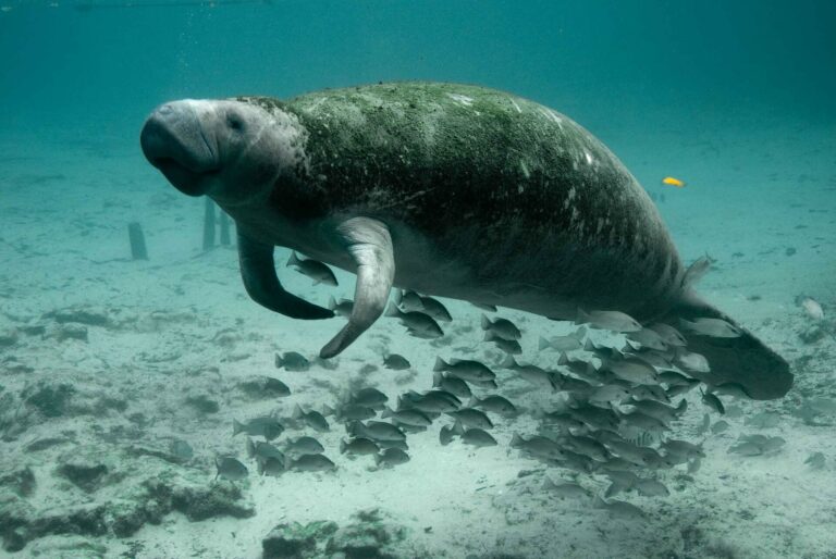
Ramos E. A., Landeo-Yauri S., Castelblanco-Martínez N., Arreola M. R., Quade A. H., Rieucau G. (2022): Drone-based photogrammetry assessments of body size and body condition of Antillean manatees. Mammalian Biology 102: 765-779.
FULL TEXT
Abstract
Assessments of individual animal health alerts to early signs of population level effects in wildlife but often rely on logistically complex wild animal captures, hindering our understanding of the wellbeing of populations in regions with limited resources. Here, we tested photogrammetry methods using small aerial drones for accurate morphometric measurements of Antillean manatee (Trichechus manatus manatus) body size and body condition. We flew drones to collect aerial imagery of captive manatees in Quintana Roo, Mexico and compared manatee body size measurements from scaled aerial imagery with physically measured body sizes. To assess optimal altitude for imaging, body size measurements acquired with an out-of-the-box drone were compared to measurements from the same drone model equipped with a LiDAR for precision altimetry flown at three altitudes (30 m, 50 m, 70 m). The accuracy of body size measures was similar for all drone models but improved with the addition of LiDAR. Difference in body size estimates between manual and drone-based measurements indicate a correction factor may be needed to account for disparities. We then used body size measurements to develop a body condition index for Antillean manatees. Our findings highlight the strength of low-cost aerial drones for morphometric measurements and assessments of manatee body condition.
PRIMATES
Breuer T., Robbins M. M., Boesch C. (2007): Using photogrammetry and color scoring to assess sexual dimorphism in wild western gorillas (Gorilla gorilla). American Journal of Physical Anthropology 134: 369-382.
FULL TEXT
Abstract
Investigating sexual dimorphism is important for our understanding of its influence on reproductive strategies including male–male competition, mate choice, and sexual conflict. Measuring physical traits in wild animals can be logistically challenging and disruptive for the animals. Therefore body size and ornament variation in wild primates have rarely been quantified. Gorillas are amongst the most sexually dimorphic and dichromatic primates. Adult males (silverbacks) possess a prominent sagittal crest, a pad of fibrous and fatty tissue on top of the head, have red crest coloration, their saddle appears silver, and they possess a silverline along their stomach. Here we measure levels of sexual dimorphism and within-male variation of body length, head size, and sexual dichromatism in a population of wild western gorillas using photogrammetry. Digital photogrammetry is a useful and precise method to measure sexual dimorphism in physical traits yielding sexual dimorphism indices (ISD), similar to those derived from traditional measurements of skeletal remains. Silverbacks were on an average 1.23 times longer in body length than adult females. Sexual dimorphism of head size was highest in measures of crest size (max ISD: 60.4) compared with measures of facial height (max ISD: 24.7). The most sexually dimorphic head size measures also showed the highest within-sex variation. We found no clear sex differences in crest coloration but there was large sexual dichromatism with high within-male variation in saddle coloration and silverline size. Further studies should examine if these sexually dimorphic traits are honest signals of competitive ability and confer an advantage in reproductive success.
Kurita H., Suzumura T., Kanchi F., Hamada Y. (2012): A photogrammetric method to evaluate nutritional status without capture in habituated free-ranging Japanese macaques (Macaca fuscata): a pilot study. Primates 53: 7-11.
FULL TEXT
Abstract
The quantification of nutritional status (e.g., total body fat) of animals is difficult, because the linear dimension (body length) required for the calculation of proxy parameters, such as the physique or body mass indices, cannot be measured without capture. One solution is photogrammetry of body length, provided the following two criteria are met: (1) the camera axes and subject are oriented vertically, and (2) anatomical landmarks are easily identified with low measurement error. By modifying Mori’s (Primates 20:371–397, 1979) approach, we devised an accurate photogrammetric method that uses a horizontal bar with an attached ruler for the monkey to traverse, and the anatomical landmarks of the eye and upper border of the ischial callosity to measure body length. We tested the applicability of this method on 11 adult female, habituated, free-ranging Japanese macaques (Macaca fuscata). Somatometric body length (crown–rump length and anterior trunk length) was statistically compared with the body length obtained using photogrammetry. The significant correlation of the photogrammetric body length with each somatometric measurement verified that the former could be employed to calculate various indices that are used to characterize fat mass (nutritional status) in Japanese macaques. The advantages and disadvantages of photogrammetry are also discussed.
Barrickman N. L., Schreier A. L., Glander K. E. (2015): Testing parallel laser image scaling for remotely measuring body dimensions on mantled howling monkeys (Alouatta palliata). American Journal of Primatology 77: 823-832.
FULL TEXT
Abstract
Body size is a fundamental variable for many studies in primate biology. However, obtaining body dimensions of wild primates through live capture is difficult and costly, so developing an alternative inexpensive and non-invasive method is crucial. Parallel laser image scaling for remotely measuring body size has been used with some success in marine and terrestrial animals, but only one arboreal primate. We further tested the efficacy of this method on the arboreal mantled howling monkey (Alouatta palliata) in La Pacifica, Costa Rica. We calculated interobserver error, as well as the method’s repeatability when measuring the same animal on different occasions. We also compared measurements obtained physically through live capture with measurements obtained remotely using parallel laser image scaling. Our results show that the different types of error for the remote technique are minimal and comparable with the error rates observed in physical methods, with the exception of some dimensions that vary depending on the animals’ posture. We conclude that parallel laser image scaling can be used to remotely obtain body dimensions if careful consideration is given to factors such as species-specific morphology and postural habits.
Galbany J., Stoinski T. S., Abavandimwe D., Breuer T., Rutkowski W., Batista N. V., Ndagijimana F., McFarlin S. C. (2016): Validation of two independent photogrammetric techniques for determining body measurements of gorillas. American Journal of Primatology 78: 418-431.
FULL TEXT
Abstract
The ability to accurately measure morphological characteristics of wild primates in the field is challenging, yet critical for understanding fundamental aspects of their biology and behavior. Recent studies have shown that digital photogrammetry can be used to non-invasively measure morphological traits of wild primates, as it allows for the determination of geometric properties of objects remotely from photographic images. We report here on a rare opportunity to test this methodology by comparing measurements obtained directly from living great apes to those obtained from photographs. We test the accuracy and precision of two independent photogrammetric techniques, employing the use of parallel lasers and a distance meter, respectively, for obtaining measurements of static objects and captive western lowland gorillas (Gorilla gorilla gorilla) (n = 4) at Zoo Atlanta. For static objects, the mean percent error between corresponding measurements collected by the same observer directly versus using photogrammetry was 0.49–0.74% for the parallel laser method and 0.62–0.76% for the distance meter method. For gorillas, mean percent error between corresponding direct and remote measurements was 2.72–5.20% for the parallel laser method and 2.20–7.51% for the distance meter method. Correlations between direct measurements and corresponding parallel laser and distance meter measurements of gorillas were highly significant with R2 values and slopes approaching 1.0 (parallel lasers: R2 = 0.9989, P < 0.0001; distance-meter: R2 = 0.9990, P < 0.0001). Further, variation between measurements of the same targets collected repeatedly by the same observer, and between different observers, was uniformly low across methods (CV, range = 0.003–0.013). While errors are slightly higher for the distance meter technique, both methods show great promise for addressing a wide range of questions requiring the non-invasive collection of morphological data from wild primates.
Galbany J., Abavandimwe D., Vakiener M., Eckardt W., Mudakikwa A., Ndagijimana F., Stoinski T. S., McFarlin S. C. (2017): Body growth and life history in wild mountain gorillas (Gorilla beringei beringei) from Volcanoes National Park, Rwanda. American Journal of Physical Anthropology 163: 570-590.
FULL TEXT
Abstract
Great apes show considerable diversity in socioecology and life history, but knowledge of their physical growth in natural settings is scarce. We characterized linear body size growth in wild mountain gorillas from Volcanoes National Park, Rwanda, a population distinguished by its extreme folivory and accelerated life histories. In 131 individuals (0.09–35.26 years), we used non-invasive parallel laser photogrammetry to measure body length, back width, arm length and two head dimensions. Nonparametric LOESS regression was used to characterize cross-sectional distance and velocity growth curves for males and females, and consider links with key life history milestones. Sex differences became evident between 8.5 and 10.0 years of age. Thereafter, female growth velocities declined, while males showed increased growth velocities until 10.0–14.5 years across dimensions. Body dimensions varied in growth; females and males reached 98% of maximum body length at 11.7 and 13.1 years, respectively. Females attained 95.3% of maximum body length by mean age at first birth. Neonates were 31% of maternal size, and doubled in size by mean weaning age. Males reached maximum body and arm length and back width before emigration, but experienced continued growth in head dimensions. While comparable data are scarce, our findings provide preliminary support for the prediction that mountain gorillas reach maximum body size at earlier ages compared to more frugivorous western gorillas. Data from other wild populations are needed to better understand comparative great ape development, and investigate links between trajectories of physical, behavioral, and reproductive maturation.
Anzà S., Berghänel A., Ostner J., Schülke O. (2022): Growth trajectories of wild Assamese macaques (Macaca assamensis) determined from parallel laser photogrammetry. Mammalian Biology 102: 1497-1511.
FULL TEXT
Abstract
Socioecological factors are associated with life-history patterns and growth trajectories among primates. Under certain conditions, selection may favor a temporal decoupling of growth and major life-history events such as sexual maturation or natal dispersal. Yet, empirical tests of these associations in wild populations remain scarce owing to the lack of non-invasive methods to capture growth trajectories. In this study, we first compared two non-invasive methods of digital photogrammetry. Then, we used parallel laser photogrammetry to investigate forearm growth of wild Assamese macaque males and females in their natural habitat at Phu Khieo Wildlife Sanctuary, Thailand to test life-history and socio-ecological hypotheses. Across 48 males and 44 females, we estimated growth trajectories and pseudo-velocity curves by applying quadratic plateau models and non-parametric LOESS regressions. We assessed the development of sexual dimorphism by comparing the sexes at five different ages. Females had completed 96% of their growth at the age at first birth (5.9 years) and ceased growing at 7.1 years of age. Males, in contrast, grew until well after their average age of natal dispersal: they reached 81% of their size at the age of natal dispersal (4.0 years), and ceased growing only at 9.0 years of age, much later than females. Sexual dimorphism in forearm length was driven by an extended growth period in males, which is expected for males dispersing between multimale and multifemale groups and not facing the risk of being ousted by other larger males. Our results contradict the neonatal investment hypothesis that predicts a desynchronization of investment in growth and reproduction only in female baboons, but not other papionins producing cheaper neonates. Furthermore, male Assamese macaques do not delay natal dispersal until they are fully grown, in accordance with predictions of the male-career-framework for species with low to medium level of direct competition.
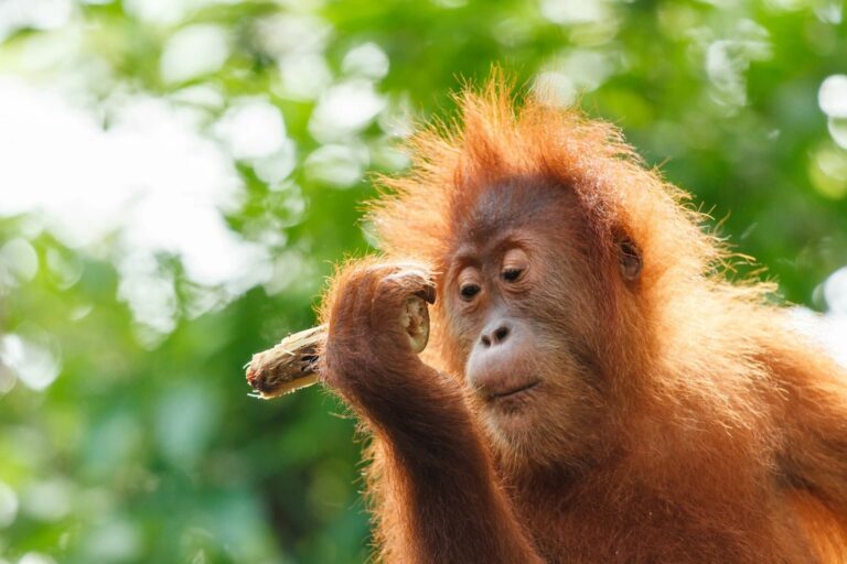
Brown E. R., Laman T. G., Kane E. E., Harwell F. S., Susanto T. W., Knott C. D. (2022): Application of a parallel laser apparatus to measure forearms and flanges of wild Bornean orangutans (Pongo pygmaeus wurmbii). American Journal of Primatology 84: e23445.
FULL TEXT
Abstract
We constructed a parallel laser photogrammetry apparatus constructed from commercially available parts, and measured forearm lengths and flange widths of 16 wild Bornean orangutans. Our objectives were to validate our method and apparatus, discuss issues encountered, and construct preliminary growth curves. For adult males, we also compared flange width to forearm length as a way to investigate the relationship between body size and flange development. We used a camera cage around a DSLR camera, on top of which we attached two parallel green lasers. We estimated error with repeatability, accuracy, and interobserver reliability measures, and measured forearm lengths in three different ways to see which was most consistent. The longest forearm measure was the most repeatable (CV = 1.64%), and was similar to flange repeatability (3.50%). Accuracy measurements of a known object were high (error = 0.25%), and Interobserver discrepancy low (3.74%). Laser spacing increased with distance to the subject, but we corrected for this using calibration photos after each session. We transparently discuss the issues we encountered with the aim that this accessible method can help expand the use of laser photogrammetry. Preliminary measurements show that male flange widths and forearm length do not reliably increase in tandem, and that female growth plateaus at around the age at first birth (15 years old). We conclude with suggested improvements to the apparatus and method to ensure the lasers remain parallel.
Richardson J. L., Levy E. J., Ranjithkumar R., Yang H., Monson E., Cronin A., Galbany J., Robbins M. M., Alberts S. C., Reeves M. E., McFarlin S. C. (2022): Automated, high-throughput image calibration for parallel-laser photogrammetry. Mammalian Biology 102: 615-627.
FULL TEXT
Abstract
Parallel-laser photogrammetry is growing in popularity as a way to collect non-invasive body size data from wild mammals. Despite its many appeals, this method requires researchers to hand-measure (i) the pixel distance between the parallel laser spots (inter-laser distance) to produce a scale within the image, and (ii) the pixel distance between the study subject’s body landmarks (inter-landmark distance). This manual effort is time-consuming and introduces human error: a researcher measuring the same image twice will rarely return the same values both times (resulting in within-observer error), as is also the case when two researchers measure the same image (resulting in between-observer error). Here, we present two independent methods that automate the inter-laser distance measurement of parallel-laser photogrammetry images. One method uses machine learning and image processing techniques in Python, and the other uses image processing techniques in ImageJ. Both of these methods reduce labor and increase precision without sacrificing accuracy. We first introduce the workflow of the two methods. Then, using two parallel-laser datasets of wild mountain gorilla and wild savannah baboon images, we validate the precision of these two automated methods relative to manual measurements and to each other. We also estimate the reduction of variation in final body size estimates in centimeters when adopting these automated methods, as these methods have no human error. Finally, we highlight the strengths of each method, suggest best practices for adopting either of them, and propose future directions for the automation of parallel-laser photogrammetry data.
Sandel A. A., Derby R. N., Chesterman N. S., McNamara A., Dudas M. M., Rawat I. (2022): Parallel lasers and digital photography to estimate limb size of chimpanzees (Pan troglodytes) at Ngogo, Kibale National Park, Uganda. Primates 63: 217-224.
FULL TEXT
Abstract
How animals grow and when they stop growing are key variables for understanding life history evolution. Although theoretically straightforward, it is logistically difficult to take body size measurements of wild animals, especially endangered and arboreal primates. Here we employ a method that has gained popularity over the past decade: digital photography combined with parallel lasers. Two laser pointers are set at a known distance apart and then projected on the animal to act as a scale in the photograph. We used this method to estimate limb length and width in a large, cross-sectional sample of mid- to late-adolescent and young adult male chimpanzees at Ngogo in Kibale National Park, Uganda. After several years of modifying our methods, we present a protocol for estimating limb length and width in wild chimpanzees. We found that by mid- to late-adolescence, male chimpanzees have reached adult height, as chimpanzees between 12 and 20 years of age did not differ in their forearm or lower leg lengths. However, mid- to late-adolescent male chimpanzees appear to continue gaining mass, as we found a weak but positive correlation between age and limb width for both forearms and lower legs. Although our method proved relatively precise, we encountered several sources of error throughout this study, such as ensuring that the lasers were indeed parallel and in identifying anatomical landmarks in the photographs. We discuss these challenges with the hope of increasing transparency and collaboration in future studies of primate body size.
Sandel A. A., Negrey J. D., Arponen M., Clark I. R., Clift J. B., Reddy R. B., Ivaska K. K. (2023): The evolution of the adolescent growth spurt: Urinary biomarkers of bone turnover in wild chimpanzees (Pan troglodytes). Journal of Human Evolution 177: 103341.
FULL TEXT
Abstract
Life history theory addresses how organisms balance development and reproduction. Mammals usually invest considerable energy into growth in infancy, and they do so incrementally less until reaching adult body size, when they shift energy to reproduction. Humans are unusual in having a long adolescence when energy is invested in both reproduction and growth, including rapid skeletal growth around puberty. Although many primates, especially in captivity, experience accelerated growth in mass around puberty, it remains unclear whether this represents skeletal growth. Without data on skeletal growth in nonhuman primates, anthropologists have often assumed the adolescent growth spurt is uniquely human, and hypotheses for its evolution have focused on other uniquely human traits. The lack of data is largely due to methodological difficulties of assessing skeletal growth in wild primates. Here, we use two urinary markers of bone turnover—osteocalcin and collagen—to study skeletal growth in a large, cross-sectional sample of wild chimpanzees (Pan troglodytes) at Ngogo, Kibale National Park, Uganda. For both bone turnover markers, we found a nonlinear effect of age, which was largely driven by males. For male chimpanzees, values for osteocalcin and collagen peaked at age 9.4 years and 10.8 years, respectively, which corresponds to early and middle adolescence. Notably, collagen values increased from 4.5 to 9 years, suggesting faster growth during early adolescence compared to late infancy. Biomarker levels plateaued at 20 years in both sexes, suggesting skeletal growth continues until then. Additional data, notably on females and infants of both sexes, are needed, as are longitudinal samples. However, our cross-sectional analysis suggests an adolescent growth spurt in the skeleton of chimpanzees, especially for males. Biologists should avoid claiming that the adolescent growth spurt is uniquely human, and hypotheses for the patterns of human growth should consider variation in our primate relatives.
UNGULATES
Willisch C. S., Marreros N., Neuhaus P. (2013): Long-distance photogrammetric trait estimation in free-ranging animals: a new approach. Mammalian Biology 78: 351-355.
FULL TEXT
Abstract
The acquisition of accurate information on the size of traits in animals is fundamental for the study of animal ecology and evolution and their management. We demonstrate how morphological traits of free-ranging animals can reliably be estimated on very large observation distances of several hundred meters by the use of ordinary digital photographic equipment and simple photogrammetric software. In our study, we estimated the length of horn annuli in free-ranging male Alpine ibex (Capra ibex) by taking already measured horn annuli of conspecifics on the same photographs as scaling units. Comparisons with hand-measured horn annuli lengths and repeatability analyses revealed a high accuracy of the photogrammetric estimates. If length estimations of specific horn annuli are based on multiple photographs measurement errors of <5.5 mm can be expected. In the current study the application of the described photogrammetric procedure increased the sample size of animals with known horn annuli length by an additional 104%. The presented photogrammetric procedure is of broad applicability and represents an easy, robust and cost-efficient method for the measuring of individuals in populations where animals are hard to capture or to approach.
Cui S., Chen D., Sun J., Chu H., Li C., Jiang Z. (2020): A simple use of camera traps for photogrammetric estimation of wild animal traits. Journal of Zoology 312: 12-20.
FULL TEXT
Abstract
The measurement of physical traits of animals is an important topic in animal ecology and wildlife management, but it is quite difficult to achieve an acceptable level of accuracy, especially for free-ranging wild animals. As a non-invasive method, camera trapping has become a common tool for detecting and monitoring wild animals. In this study, we combined camera-trapping images and a photogrammetry technique to present a camera trap-based photogrammetric estimation (CTPE) approach for robust measurement of morphological traits of animals. We first tested our method in a captive population of fallow deer (Dama dama) and compared the estimated shoulder heights by CTPE with corresponding hand measurements. The results revealed a remarkable accuracy, with an expected measurement error within ± 5 cm. Second, we mainly demonstrated a field application of CTPE in a wild population of Altai wapiti (Cervus elaphus sibiricus) in the Kanas National Nature Reserve, Xinjiang, China. For wapiti, we found that the average shoulder height of males was 135 ± 9 cm (mean ± sd, n = 30) and that of females was 125 ± 6 cm (n = 37), with all data significantly fitting a normal distribution. We did not find any differences in CTPE results among cameras, between photograph types (colour vs. black and white) or temperatures. No significant difference existed between our results and the hand-measured shoulder heights of wapiti from deer farms. Finally, we discussed the strengths, limitations and potential extensions of the method. We suggest that the combination of camera trapping and the photogrammetry technique is quite promising and that CTPE has great potential in wildlife field investigations.
Martin J. M., Barboza P. S. (2020): Decadal heat and drought drive body size of North American bison (Bison bison) along the Great Plains. Ecology and Evolution 10: 336-349.
FULL TEXT
Abstract
Large grazers are visible and valuable indicators of the effects of projected changes in temperature and drought on grasslands. The grasslands of the Great Plains have supported the greatest number of bison (Bison bison; Linnaeus, 1758) since prehistoric times. We tested the hypothesis that body mass (BM, kg) and asymptotic body mass (ABM, kg) of Bison decline with rising temperature and increasing drought over both temporal and spatial scales along the Great Plains. Temporally, we modeled the relationship of annual measures of BM and height (H, m) of 5,781 Bison at Wind Cave National Park (WICA) from 1966 to 2015. We used Gompertz equations of BM against age to estimate ABM in decadal cohorts; both females and males decreased from the 1960s to the 2010s. Male ABM was variable but consistently larger (699 vs. 441 kg) than female ABM. We used local mean decadal temperature (MDT) and local mean decadal Palmer Drought Severity Index (dPDSI) to model the effects of climate on ABM. Drought decreased ABM temporally (−16 kg/local dPDSI) at WICA. Spatially, we used photogrammetry to measure body height (HE) of 773 Bison to estimate BME in 19 herds from Saskatchewan to Texas, including WICA. Drought also decreased ABM spatially (−16 kg/local dPDSI) along the Great Plains. Temperature decreased ABM both temporally at WICA (−115 kg/°C local MDT) and spatially (−1 kg/°C local MDT) along the Great Plains. Our data indicate that temperature and drought drive Bison ABM presumably by affecting seasonal mass gain. Bison body size is likely to decline over the next five decades throughout the Great Plains due to projected increases in temperatures and both the frequency and intensity of drought.
Smith A. F., Bongi P., Ciuti S. (2021): Remote, non‐invasive photogrammetry for measuring physical traits in wildlife. Journal of Zoology 313: 250-262.
FULL TEXT
Abstract
Morphometric measurements in wildlife are essential data needed to link behaviour and ecology to life-history traits. However, acquisition of such data, especially in large vertebrates, is a challenging and limiting aspect in many studies due to associated capture and handling. Ultimately, this prevents testing important hypotheses in wildlife ecology. We demonstrate how to overcome some of these challenges by improving the far-reaching potential of photogrammetry for wildlife. We validate a methodology using photogrammetry to acquire measurements of complex, curvilinear, morphometric traits (deer antlers) at long range (up to 200 m), providing a methodological foundation for further application to numerous physical attributes and species. We calibrated and validated two different photogrammetric camera systems and applied long-range photogrammetry to measure a suite of antler features from deer trophies positioned analogous to free-living, wild deer. We also developed, validated and described an efficient correction protocol to overcome the complexity of three-dimensional antler features. This complexity previously biased the acquisition of absolute measurements. Our measurement estimates were characterized by mean errors lower than 5% when compared to true sizes, with our correction protocol for antlers particularly effective and validated with independent antler sizes. Importantly, the estimated size rankings based on the true sizes were identical to the rankings estimated by photogrammetry in all cases. We measured antlers in free-living, wild fallow deer as a field validation and demonstration, showing how we were able to collect physical trait measurements of 230 antlers without capture. This non-invasive, remote and fast technique can provide valuable and accurate data allowing researchers to address new hypotheses by linking life-history traits to wildlife ecology, behaviour and management.
Gherissi D. E., Lamraoui R., Chacha F., Gaouar S. B. S. (2022): Accuracy of image analysis for linear zoometric measurements in dromedary camels. Tropical Animal Health and Production 54: 232.
FULL TEXT
Abstract
The present study was designed to verify the effectiveness of the image analysis method for body measurement in dromedary camel compared to manual measurements as a reference method. To achieve this aim, twenty-one linear body measurements were estimated on 59 adult Sahraoui dromedary camels (22 males and 37 females) with a normal clinical condition by using a measuring stick or vernier caliper (standard method). On the other hand, image analysis on profile, front, or behind photographs was processed using Axiovision Software. Overall mean comparison, relative error, variance, Pearson’s correlation coefficient, and coefficient of variance showed that the image analysis method was accurate in relation to the manual measurement. Furthermore, image analysis results indicated relevant accuracy (bias correction factor, Cb ≈1) and precision (Pearson ρ ≈1) which were significantly correlated with the results of the reference method (Lin’s concordance correlation coefficients rccc ≈ 1). According to Bland–Altman upper and lower limits of agreement, the concordance was estimated between 93.22 and 98.3%. Passing-Bablok regression showed a good relationship between the results of the two methods displaying no significant systematic and proportional bias. The image analysis method for linear body measurements in dromedary camel showed results that are in agreement with the manual measuring method. Therefore, the image analysis could be considered a valid tool for camel conformation trait studies.
Inman V. L., Leggett K. E. (2022): Hidden hippos: Using photogrammetry and multiple imputation to determine the age, sex, and body condition of an animal often partially submerged. Drones 6: 409.
FULL TEXT
Abstract
Demographic Information on threatened species is important to plan conservation actions. Due to their aquatic lifestyle, the subtle nature of hippo sexual dimorphism, and their occurrence in inaccessible areas, it is difficult to visually determine hippo ages and sexes. Previously, hippo body lengths have been measured from drone images and used to estimate age. However, due to hippos’ propensity to be partially submerged, it is often difficult to obtain the required measurements. We used the novel technique of multiple imputation to estimate missing body measurements. Further, we explored if male and female hippos could be differentiated in drone images based on body proportions, also examining body condition indices and how these varied seasonally. Multiple imputation increased the number of hippos that we aged threefold, and the body lengths we obtained fell within the range provided in literature, supporting their validity. We provide one of the first age structure breakdowns of a hippo population not from culled hippos. Accounting for overall size, males had wider necks and snouts than females. Hippo body condition varied seasonally, indicating responses to resources and reproduction. We provide a new technique and demonstrate the utility of drones to determine age and sex structures of hippo populations.
Leorna S., Brinkman T., Fullman T. (2022): Estimating animal size or distance in camera trap images: Photogrammetry using the pinhole camera model. Methods in Ecology and Evolution 13: 1707-1718.
FULL TEXT
Abstract
As camera trapping has become a standard practice in wildlife ecology, developing techniques to extract additional information from images will increase the utility of generated data. Despite rapid advancements in camera trapping practices, methods for estimating animal size or distance from the camera using captured images have not been standardized. Deriving animal sizes directly from images creates opportunities to collect wildlife metrics such as growth rates or changes in body condition. Distances to animals may be used to quantify important aspects of sampling design such as the effective area sampled or distribution of animals in the camera’s field-of-view. We present a method of using pixel measurements in an image to estimate animal size or distance from the camera using a conceptual model in photogrammetry known as the ‘pinhole camera model’. We evaluated the performance of this approach both using stationary three-dimensional animal targets and in a field setting using live captive reindeer Rangifer tarandus ranging in size and distance from the camera. We found total mean relative error of estimated animal sizes or distances from the cameras in our simulation was −3.0% and 3.3% and in our field setting was −8.6% and 10.5%, respectively. In our simulation, mean relative error of size or distance estimates were not statistically different between image settings within camera models, between camera models or between the measured dimension used in calculations. We provide recommendations for applying the pinhole camera model in a wildlife camera trapping context. Our approach of using the pinhole camera model to estimate animal size or distance from the camera produced robust estimates using a single image while remaining easy to implement and generalizable to different camera trap models and installations, thus enhancing its utility for a variety of camera trap applications and expanding opportunities to use camera trap images in novel ways.
Kido N., Tanaka S., Wada Y., Oura A., Ochiai E., Morita N., Kawaguchi Y., Itabashi M., Munakata T. (2024): Body shape analysis in reticulated giraffe, okapi, and black rhinoceros using three-dimensional laser measurements. Journal of Zoological and Botanical Gardens 5: 80-89.
FULL TEXT
Abstract
Health management in captive animals is difficult for various unknown reasons presumably related to physiological functions, disease, and diet. Generally, abnormal conditions are diagnosed based on body weight; however, zoos lack appropriate scales for megafauna. Body shape evaluation is often used to evaluate the nutritional status of breeding animals; however, this is inaccurate for zoo animals because of inter-observer variability, especially in megafauna. Previously, three-dimensional laser measurements were used to analyse body shape of reticulated giraffe, but further studies are required to examine its effectiveness in more individuals, and other species. Here, we applied this method to seven reticulated giraffe (Giraffa reticulata), five okapi (Okapia johnstoni), and three black rhinoceros (Diceros bicornis) individuals for which cross-sectional area, width, and height in transverse section were determined. Relative change rates of each variable in relation to measurements at the axillary region revealed changes in body shape for each individual. Further, scatter plots and corresponding fitted curves and correlation coefficients showed a correlation between body length and approximate volume. The accuracy of three-dimensional laser measurements was demonstrated in three animal species, whereby we propose its use as an alternative method to evaluate body shape in megafauna without the inter-observer variability. In addition, this handheld device may be applied for various zoos without the scale for megafauna.


