MORPHOLOGY: MOLLUSCS
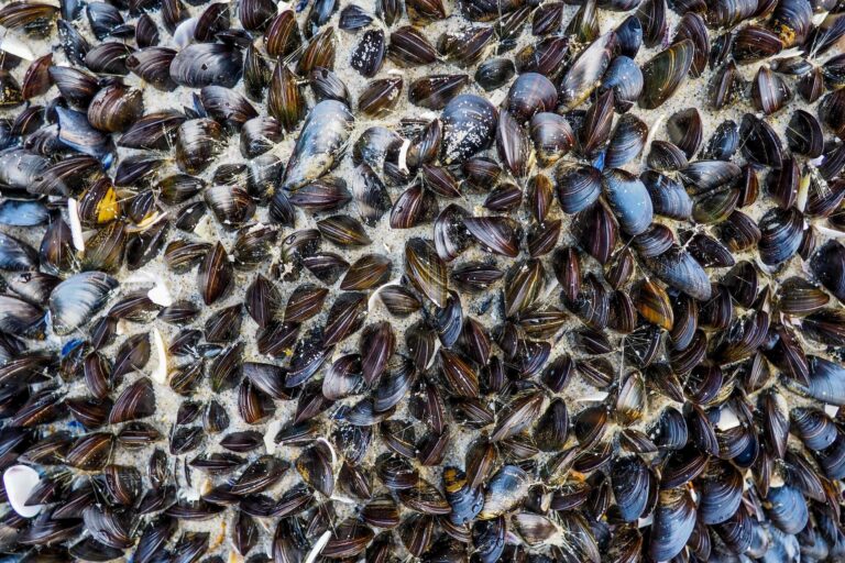
BIVALVES
Wilson C. D., Jane Preston S., Moorkens E., Dick J. T., Lundy M. G. (2012): Applying morphometrics to choose optimal captive brood stock for an endangered species: a case study using the freshwater pearl mussel, Margaritifera margaritifera (L.). Aquatic Conservation: Marine and Freshwater Ecosystems 22: 569-576.
FULL TEXT
Abstract
To maximize captive breeding success for the globally endangered freshwater mussel Margaritifera margaritifera, morphometrics was applied to develop a tool for selecting optimal brood stock. There was high discrimination between brooding and non-brooding individuals and the presence of brood explained the variation in the percentage of mussels with a typical brooding morphology. Brooding individuals were significantly wider than non-brooding individuals. However, after reclassifying those non-brooding individuals with morphology highly indicative of brooding individuals using Mahalanobis distance modelling, only shell curvature along the ventral region differed significantly. The Mahalanobis model explained more variation in shell morphology than a model based on field observations, highlighting that shell morphology is a good predictor of brooding mussels. In addition, it could be argued that an identified novel morph is that of hermaphroditic M. margaritifera, which has developed in response to historic low population density. This is the first application of a non-invasive, morphometric technique to optimize captive breeding programmes for an endangered species. Since a greater number of species are under threat of extinction from climate change, there will be a demand for captive breeding programmes, emphasizing the importance of this study.
Cordero-Rivera A., Ondina P., Outeiro A., Amaro R., San Miguel E. (2022): Allometry in the freshwater pearl mussel (Margaritifera margaritifera L.): Mussels tend to grow flatter at higher water speed. Malacologia 64: 257-267.
FULL TEXT
Abstract
The freshwater pearl mussel (Margaritifera margaritifera) is one of the longest-lived invertebrate species in the world and one of the most threatened freshwater animals in Europe. Its southernmost populations, located in northwestern Spain, are in a critical conservation situation and are still understudied. Here we calibrate a non-invasive method for calculating the volume of the shell and use it to study the ontogenetic scaling of shell volume on shell length. We characterized ontogenetic growth and determined allometric relationships in 16 M. margaritifera northwestern Spain populations by using ordinary least squares regression, major axis and reduced major axis methods. We estimated topographic slopes of the sampling points using a GIS system, as a proxy of water speed. We measured 803 shells and found that the volume of the shell can precisely be estimated using three linear measurements. We found evidence for negative allometry of shell volume in the global sample and in 11 populations. We hypothesized that water speed would affect allometric patterns of local populations. Results suggest a negative relationship between the allometric slope and the topographic slope of the river section inhabited by M. margaritifera. We propose that when water speed is higher, larger mussels become proportionally flatter than in locations where water current is slower, allowing them to burrow more easily in the sediment. Our method will allow estimation of M. margaritifera biomass and ontogenetic growth without killing any specimens, which will contribute to conservation programs for this species.
Pineda‐Metz S. E., Merk V., Pogoda B. (2023): A machine learning model and biometric transformations to facilitate European oyster monitoring. Aquatic Conservation: Marine and Freshwater Ecosystems.
FULL TEXT
Abstract
Ecosystem monitoring, especially in the context of marine conservation and management requires abundance and biomass metrics, condition indices, and measures of ecosystem services of key species, all of which can be calculated using biometric transformation factors. Following ecosystem restoration measures in the North Sea and north-east Atlantic waters, European oyster (Ostrea edulis) restoration and its monitoring have substantially increased over the past decade. Restoration activities are implemented by diverse approaches and practitioners ranging from governmental conservation agencies, research institutions and non-governmental institutions to regional groups, including citizen science projects. Thus, tools for facilitating data acquisition and estimation with non-destructive techniques can support monitoring quantitatively and qualitatively. Weight-to-weight transformation factors for calculating dry weight of O. edulis from wet weight measurements are presented. Another important tool is the estimation of weight only from size measurements. The classical approach to achieve these transformation factors is the construction of allometric models, which, however, can greatly vary among regions and between years, making them extremely location/season specific. Alternative and more flexible models constructed using random forests are proposed. This algorithm is a machine learning technique that is increasingly used in ecology, and has been proven to outperform other predictive models. From biometric variable measurements of 1,401 O. edulis individuals, allometric models were used to estimate total, shell and body wet weights, and compare them with 15 random forest models. In general, the random forest models outperformed the allometric ones, with lower error when estimating weight. The developed random forest models can thus provide a tool for facilitating oyster restoration monitoring by increasing data acquisition without the need of sacrificing European oyster individuals. Their improvement can imply its implementation in other regions and support European oyster restoration and monitoring efforts throughout Europe.
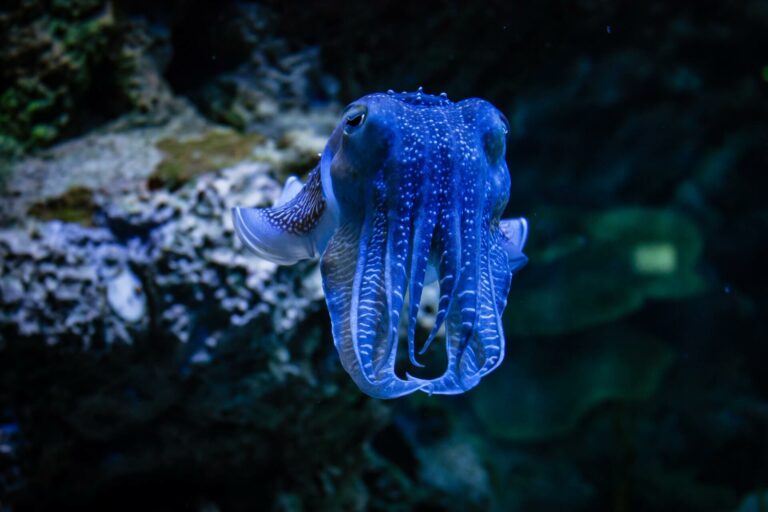
CEPHALOPODS
Ziegler A., Sagorny C. (2021): Holistic description of new deep sea megafauna (Cephalopoda: Cirrata) using a minimally invasive approach. BMC Biology 19: 81.
FULL TEXT
Abstract
In zoology, species descriptions conventionally rely on invasive morphological techniques, frequently leading to damage of the specimens and thus only a partial understanding of their structural complexity. More recently, non-destructive imaging techniques have successfully been used to describe smaller fauna, but this approach has so far not been applied to identify or describe larger animal species. Here, we present a combination of entirely non-invasive as well as minimally invasive methods that permit taxonomic descriptions of large zoological specimens in a more comprehensive manner. Using the single available representative of an allegedly novel species of deep-sea cephalopod (Mollusca: Cephalopoda), digital photography, standardized external measurements, high-field magnetic resonance imaging, micro-computed tomography, and DNA barcoding were combined to gather all morphological and molecular characters relevant for a full species description. The results show that this specimen belongs to the cirrate octopod (Octopoda: Cirrata) genus Grimpoteuthis Robson, 1932. Based on the number of suckers, position of web nodules, cirrus length, presence of a radula, and various shell characters, the specimen is designated as the holotype of a new species of dumbo octopus, G. imperator sp. nov. The digital nature of the acquired data permits a seamless online deposition of raw as well as derived morphological and molecular datasets in publicly accessible repositories. Using high-resolution, non-invasive imaging systems intended for the analysis of larger biological objects, all external as well as internal morphological character states relevant for the identification of a new megafaunal species were obtained. Potentially harmful effects on this unique deep-sea cephalopod specimen were avoided by scanning the fixed animal without admixture of a contrast agent. Additional support for the taxonomic placement of the new dumbo octopus species was obtained through DNA barcoding, further underlining the importance of combining morphological and molecular datasets for a holistic description of zoological specimens.
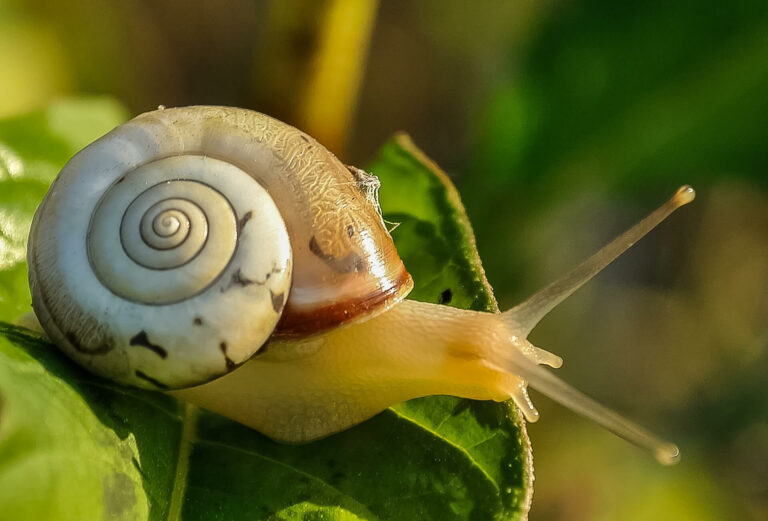
GASTROPODS
Perea J., Garcia A., Acero R., Valerio D., Gómez G. (2008): A photogrammetric methodology for size measurements: application to the study of weight–shell diameter relationship in juvenile Cantareus aspersus snails. Journal of Molluscan Studies 74: 209-213.
FULL TEXT
Abstract
A photogrammetric methodology is proposed to measure the diameter of snail shells on digital pictures. Digital photographs were taken of juvenile Cantereus aspersus snails. AutoCAD image analysis software was used for the measurements and the results were contrasted with data obtained using a digital caliper in the same snails to compare the accuracy of both methods. The snails were individually weighed and the shell diameter was measured once a week, for a total of 7 weeks. After the third week, there were no significant differences between both methods, whereas in the first 2 weeks the measurements obtained with a digital caliper scored larger diameters than the photogrammetric measurements. This is probably due to the difficulty to define the end of the shell with the caliper, whereas the photogrammetric analysis does not involve any risk for the snail. To test the reproducibility and repeatability of both methodologies seven snails were measured five times by three different examiners. Using the variance components analysis, the repeatability was 4.8% of the total variation for the photogrammetric methodology and 6.9% for the conventional methodology, while the reproducibility was 0.0% and 2.7% for the photogrammetric and conventional methodology, respectively. These findings indicate that the photogrammetric methodology can be used with confidence to measure the diameter of snail shells. The great advantage of this method is the ability to magnify the images to make more precise measurements from snails of any size, which is especially useful in the early stages of development. The growth data were used to construct a model for live weight (LW) estimation based on shell diameter. The snails showed high growth rates both in terms of shell diameter and LW. The shell diameters showed a low individual variation (16.2% CV) and were normally distributed, whereas the LWs showed greater variability (36.8% CV) and were not normally distributed. The best model for estimating LW from shell diameter in juvenile snails is the multiplicative model, with a high determination coefficient (96%).
Irniger A., Morari M. J., Bürkli A., Detert M. (2017): Automatic high-throughput measurement of live aquatic snails from images. Journal of Molluscan Studies 83: 235-239.
FULL TEXT
Excerpt
We developed and tested an algorithm that enables the automatic, noninvasive detection and measurement of living aquatic snails that are too small and too numerous for conventional measuring methods. This algorithm allows simultaneous measuring of the area, length and width of multiple snails per image.
Beindorff N., Schmitz-Peiffer F., Messroghli D., Brenner W., Eary J. F. (2021): Radionuclide, magnetic resonance and computed tomography imaging in European round back slugs (Arionidae) and leopard slugs (Limacidae). Scientific Reports 11: 13798.
FULL TEXT
Abstract
Other than in animal models of human disease, little functional imaging has been performed in most of the animal world. The aim of this study was to explore the functional anatomy of the European round back slug (Arionidae) and leopard slug (Limacidae) and to establish an imaging protocol for comparative species study. Radionuclide images with single photon emission computed tomography (SPECT) and positron emission tomography (PET) were obtained after injections of standard clinical radiopharmaceuticals 99mtechnetium dicarboxypropane diphosphonate (bone scintigraphy), 99mtechnetium mercaptoacetyltriglycine (kidney function), 99mtechnetium diethylenetriaminepentaacetic acid (kidney function), 99mtechnetium pertechnetate (mediated by the sodium-iodide symporter), 99mtechnetium sestamibi (cardiac scintigraphy) or 18F-fluoro-deoxyglucose (glucose metabolism) in combination with magnetic resonance imaging (MRI) and computed tomography (CT) for uptake anatomic definition. Images were compared with anatomic drawings for the Arionidae species. Additionally, organ uptake data was determined for a description of slug functional anatomy in comparison to human tracer biodistribution patterns identifying the heart, the open circulatory anatomy, calcified shell remnant, renal structure (nephridium), liver (digestive gland) and intestine. The results show the detailed functional anatomy of Arionidae and Limacidae, and describe an in vivo whole-body imaging procedure for invertebrate species.


